RIFE CRANE |

|
|
|
|
A PHYSICIST’S
VIEW OF DR. RIFE’S NON-DRUG TREATMENT (This is a revised and updated version of an article, which
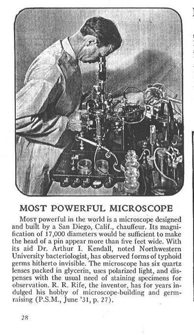
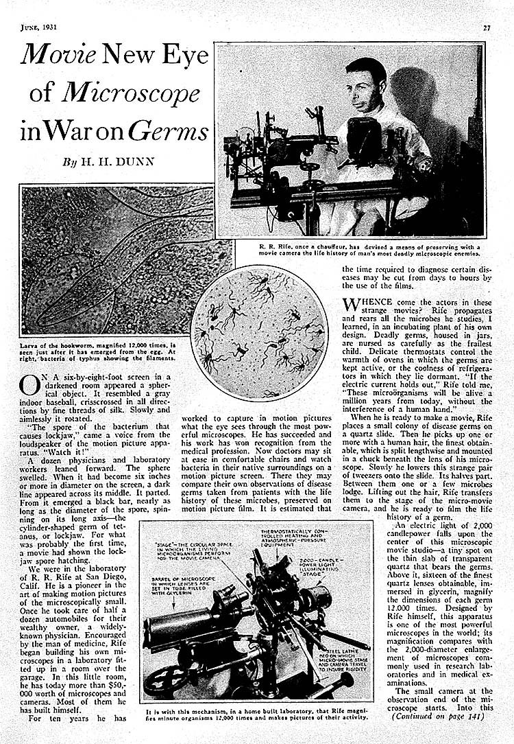
was originally released in the First, he invented a new kind of optical microscope. This microscope could be used to observe viruses in live cells and tissue culture. (1,2) Rife built five of these microscopes. Rife never published the plans to his microscope and to this day those in the scientific establishment not familiar with Rife’s work wrongly claim that it is generally impossible to see or identify a virus with any optical microscope (see Rife microscope addendum). Rife’s second great accomplishment was to invent a variable frequency flashing light ultrasound source which could kill bacteria, rickettsias, protozoa, fungi, and viruses. By 1939, Dr. Rife had both identified the microbes and light flashing rates ( ultrasound frequencies ) required to kill these microbes, which were associated with fifty-two major diseases, including carcinoma and sarcoma cancers. (2,3) In this article we will describe what Rife’s frequency instrument was and how it could destroy a microbe without harming the host / patient. Figure 1 shows a schematic view of the Rife frequency instrument in operation treating a patient. The frequency instrument consisted of an old style X-ray tube, which had been back filled with helium and or argon gas at very low pressure and had a current flow through the tube driven by a packetted sine and or square wave voltage oscillations as depicted in Figure 2. Each time the plate-anode voltage polarities reverse as depicted in Figure 2, there is an associated current surge reversal and shock wave generation in the tube gas. The shock wave travels to the tube wall and vibrates it producing sound / ultrasound in the air at the tube outer surface. The electron current collides with the tube gas optically exciting and ionizing the gas atoms. The instantaneous light intensity output of the tube is approximately proportional to the instantaneous tube current plotted in Figure 3. Therefore, the plot of the light intensity from the tube gas discharge verses time will have the same shape as that of Figure 3. Note that every other current and or light pulse has a different peak amplitude. This difference in current / light intensity amplitude is do to the preferential electron current flow from the hot tungsten cathode to the metal plate anode. The hot cathode when at a negative polarity (voltage) relative to the anode readily emits electrons for acceleration across the tube. However, when the “anode” is at a negative voltage relative to the hot tungsten cathode, the electron current across the tube must build up from electrons released by positive ions colliding with the plate, electrons emitted by the plate from photo-emission caused by ultraviolet light from meta-stable atoms, and accelerated electrons colliding with atoms and freeing new electrons to join the current flow. Note that there are two shock wave pulses per single electrical oscillation cycle. Also, note there are two light pulses per single electrical oscillation. In other words there is a frequency doubling effect, i.e. a one million cycle per second sine wave voltage across the Rife tube will produce ultrasound with strong components at two million cycles per second. As a practical example, Rife found that the common carcinoma breast cancer of his time, (which is now reaching epidemic proportions among women), was killed by packetted sine and or square wave voltages in the Rife tube of a frequency of 11,780,000 cycles per second. This means light flashing rates and ultrasound of 23,560,000 cycles per second will be produced by Rife’s tube.(4,5) For the currently trained biologist and medical researcher, all of the above statements about Rife’s work and accomplishments are suspect at best. The reason for this is that they have a very limited knowledge of physics and no knowledge of Rife’s research results. For example, they do not know that Rife isolated a viral sized, spore like, motile form of the E-coli bacteria that when exposed to prolonged ultraviolet light became a virulent carcinogen, which invariably caused carcinoma cancer when injected into lab animals. The key to getting Rife’s work and accomplishments into general medical and biological use is an end to this ignorance about how and why Rife’s frequency instrument worked to kill microbes. To that end I will now describe and illustrate how the Rife frequency instrument can destroy a virus. I will illustrate how a specific virus can be destroyed by a specific frequency of ultrasound. This ultrasound is generated by the Rife frequency instrument by three methods. In the first method, note that light carries linear momentum and that when the pulse of light from the Rife frequency instrument is absorbed and or reflected by the patient’s skin layer that skin layer must recoil in the direction of light flow from the tube to conserve linear momentum. When the light pulse has ended, the skin relaxes back toward its non-light pulse exposure position. In other words, periodic light pulses generate periodic pressure pulses in the patient’s skin layer, which travel into the patient’s body. The Rife frequency instrument converts the patient’s entire exposed skin surface into an ultrasound transducer for the generation of ultrasound. Even though the efficiency of ultrasound production is exceedingly low by this method, it is still adequate to kill microbes, because we are dealing here with a resonance phenomenon. In the second method, the plasma shock waves inside the Rife tube vibrate the tube wall causing generation of ultrasound in the air. This ultrasound travels to and enters the patient. In the third method, the oscillating electric fields from the Rife tube travel out into the room and interact with (vibrate) the charged ions in the patient’s body. These vibrating ions generate ultrasound in the patient of the same frequency as the electric field vibration rate of the Rife ray tube (no frequency doubling effect). As a practical example, Rife would treat his cancer patients using his frequency instrument for three minutes of exposure once every three days. Usually his “terminally” ill cancer patients would be cancer free in about thirty such treatments or less, as was verified in the 1934, 1935, and 1937 test clinical trials carried out by the U.S.C. Medical School Special Medical Research Committee. (3) That same committee then suppressed the research results. The reason for the short three minute treatment is to kill off only a thin outer layer of cancer tumor tissue at one time. This allows the body’s immune system to remove this layer before the next treatment. The entire cancer tumor could have been killed / destroyed in a single Rife frequency instrument treatment of perhaps one to one and a half hours. However, then the cancer patient would have a large mass or masses of dead cancer tissue in them, which would become a feast for a massive bacterial infection. This bacterial infection could lead to liver and kidney damage and general toxemia. The Rife frequency instrument killed the “normal” carcinoma cancer cell of Rife’s time by rupturing the thousands of BX cancer viruses they contain and thereby dumping the BX cancer virus contents into the cancer cell cytoplasm. This BX cancer virus as Rife named it in 1931 is not a virus by the normal standard usage of the term virus today. Rife based his definition on the fact that the BX cancer virus could pass through the finest Berkett porcelain filter of the time (000 filter). The BX cancer virus is ovoid in shape, .066 microns along the major axis and .05 microns along the minor axis. It is motile, driven by a proton transport flagella the same as its bacterial parent, the E-coli bacteria. When this BX cancer virus is ruptured it spills out its gnome, ribosomes, RNA, enzymes, and various proteins. When thousands of these ruptures occur all at once in a carcinoma cancer cell the results are fatal to the cancer cell. A similar situation occurs in the sarcoma cancer cell when the BY cancer viruses are all disintegrated at once, The BY cancer virus is another form of the BX cancer virus which Rife found caused sarcoma cancer after it had been exposed to prolonged ultraviolet light exposure. To see how the ultrasound generated by the Rife frequency instrument can destroy a virus we will examine the outer protein coat (capsid) structure of a virus. Most viruses of interest which cause disease in plants and animals have an icosahedral capsid structure as illustrated in Figure 4A and B. A specific example of this icosahedral capsid structure is illustrated in Figure 5. Each dark circle represents a spherical protein molecule clump. When the virus capsid of Figure 5 is folded together as indicated in Figure 4A and B, a simple virus capsid model has been formed. Examination of this capsid model shows a large number of intersecting and overlapping closed rings of protein molecule clumps. These closed rings of periodically spaced protein clumps are illustrated in Figure 6A and B. In classical physics when studying standing wave phenomenon the periodically spaced protein clumps, as illustrated in Figure 6A and B, are known as the mass beads on a string with circular boundary conditions problem. Figures 7A, B and C, illustrated this classical physics problem. Figures 8A, B, C, and D illustrate some of the standing wave motion modes which the closed periodically spaced protein clump rings of Figure 6A can sustain. Figure 8A shows a ten member protein clump ring linearized for ease of graphing wave motion displacement of the center of the protein clumps from their equilibrium position. Figure 8B shows the most stressful oscillation mode for the ten member protein clump ring. In this oscillation mode, adjacent protein molecule clumps are always going in opposite directions and therefore putting maximum stress on where they are bonded together. If this oscillation mode is raised to a high enough displacement amplitude the ring will rupture, If enough rings are ruptured, the virus capsid disintegrates. The Rife frequency instrument when set to the frequency which corresponds to the most stressful oscillation mode for the virus of interest, as illustrated in Figure 8B, will destroy that virus capsid coat and therefore destroy the virus. The common virus capsid coat was chosen to show how the Rife frequency instrument could destroy a microbe which has closed on themselves periodically spaced protein clump structures.(6) Bacteria, protozoa, rickettsias, and fungi all have versions of these closed on themselves periodically spaced protein clump structures, in their structure, which makes them susceptible to destruction by the Rife frequency instrument (specific frequency of ultrasound). Advancements in electron technology have made it possible to obtain Rife frequency instrument results (generation of specific frequencies of ultrasound) using several different approaches. For example, electrodes applying a square wave voltage to the bare skin, oscillating capacitively coupled high voltage discharges through gas discharge tubes touching the bare skin, multi wave multipole electromagnetic radiation devices, intense very rapidly changing (pulsed) in strength magnet field devises, and a piezo-electric transducer driven by an appropriate voltage signal source. In our present circumstances where antibiotic resistant bacteria are about to become rampant, anti-viral drugs are largely still just a bio-tech dream, and the war on cancer has been a dismal failure for the cancer patient, but not for the so-called cancer researcher. It is long since time for Rife's 1930,s work to be implemented. Final note: When looking at Rife’s actual lab note book for the mortal oscillation rate for a particular microbe, you find a frequency and a wave length listed for that microbe.(ref. 7) However, further examination of the note book shows that the given frequency and wave length are not related to each other. In other words the product of the wave length times the frequency listed for various microbes does not give a constant value for wave propagation. The logical conclusion is that Rife was using two frequency of electrical oscillation in his frequency instrument. One frequency Rife gave directly and the other is implied by the wave length given, because when you have a wave length you always have an associated frequency from the fundamental relationship that wave length multiplied by the associated frequency equals wave propagation speed. When using two different frequencies of electrical oscillations in an amplifier circuit which Rife used in his frequency instrument, there are two obvious possibilities of what can be done: 1) One oscillation frequency can be used to amplitude modulate the other, and 2) The two oscillation frequencies can be added together and then amplified. This article has assumed the first possibility. If I had of assumed the second possibility, then qualitatively the results would be the same except that there would now be a new additional strong ultrasound frequency component generated by the tube of a frequency equal to the difference between the two frequencies Rife used. This situation is illustrated in Figures 9A and B. I have attempted to verify the validity of this second possibility using “known” mortal oscillation rates for specific microbes obtained from correspondence between Rife and his associates. However, so far all my calculated results are contradictory. REFERENCES: 1) The New Microscopes, by R.E. Seidel, M.D. and
M. Elizabeth Winter, Journal of The Franklin Institute, Vol. 237, Feb.
1944.
|