RIFE CRANE |

|
|
|
|
Proceedings Published Weekly for the Information of the Members of the Staff and the Fellows of The Mayo Foundation for Medical Education and Research -------------------------------------------------------------------------------- Vol. 7 Rochester, Minnesota, Wednesday, July 13, 1932 No.28
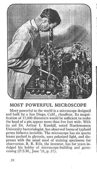
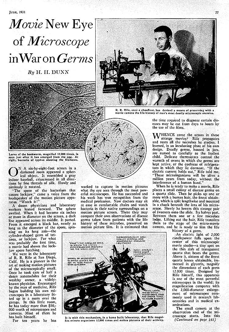
It is the purpose of the report to record the more important observations made during three days, July 5 6, and 7, 1932, spent with them in Dr. Ken all’s laboratory at Northwestern University Medical School, Chicago. Owing to the novel and important character of the work, each of us verified at every step the results obtained. Microscopic examination of suitable specimens was made as a routine by Dr. Rife with his high-power microscope, by Dr. Kendall with the oil immersion dark field, and by myself with the ordinary Zeiss microscope equipped with a 2 mm. apochromatic oil immersion lens and x10 ocular giving a magnification of about 900 diameters. Most obesrvations with the Rife microscope were made at 8,000 diameters. In order to check the magnification, gram and safranin stained films of cultures of Eberthella typhi, of the streptococcus from poliomyelitis, and stained films of blood, and of the sediment of the spinal fluid from a case of acute poliomyelitis, were examined. Bacilli, streptococci, erythrocytes, polymorphonuclear leukocytes, and lymphocytes were clearly seen, and in each instance were, as near as could be estimated, about nine times the diameter as when examined with the 2 mm. oil immersion at about 900 diameters. The following principles and methods were stated by Dr. Rife as being essential in order to visualize clearly the objects at this and higher magnification by direct observation. Spherical aberration is reduced to the minimum and magnification greatly increased by using objectives in place of oculars. Proper visualization, especially of unstained objects, is obtained by the use of an intense beam of monochromatic polarized light created by rotating wedge-shaped quartz prisms placed between the source of light and the substage quartz condensor. Dispersion of the transmitted rays of light, as they pass upward to the eye, is prevented by passing them through a series of quartz erecting (90 deg) prisms. Projection of the rays of light through air is not greater than 30 mm. at any point. The oval, motile, turquoise blue bodies described previously by Kendall and Rife were demonstrated unmistakably under the Rife microscope in numerous hanging drop, between slide and cover glass, preparations of old and young cultures and corresponding filtrates of two strains of Eberthella typhi. One of these strains represented the one studied by them previously, the other was isolated a short time before from the blood in a case of typhoid fever. The bodies were numerous in cultures of the bacilli in the K medium but were also found in smaller numbers in peptone broth cultures. The motile forms, as a rule, were of a deeper blue-green color than nonmotile forms. In several instances they were seen attached to, or were in the swollen ends of, bacilli having a similar turquoise blue color. In one instance, the turquoise blue body was seen to be extruded or detached and as this occurred the bacillus lust its bluish-green color. The bacilli not containing granules seemed to be of different density and were of a grayish-brown color instead. No bacilli were found in clouded cultures in K medium inoculated previously with filtrates of cultures; only large numbers of the bluish-green bodies were seen. This was true of the strain examined last November and which has been kept in filtrable, nonbacillary form through a series of transfers ever since, as well as their recently isolated strain. Stained and hanging drop preparations of clouded filtrate cultures examined under my microscope were uniformly negative, despite the fact that the blue bodies, as seen under the Rife microscope, appeared not less in diameter than the cross-diameter of the bacilli. Dancing bodies were seen under dark field illumination. These were considered as the filtrable, turquoise blue bodies. The turquoise blue bodies were not found in filtrates of the uninoculated K medium. Bodies resembling those of Eherthella typhi, but of grayish-brown color were seen in K medium cultures of Eschericbia coli and corresponding filtrates. It was next agreed to filter cultures of the streptococcus from poliomyelitis which I brought with me, for the purpose of controlling the results obtained with Eberthella typhi, as well as to see what the Rife microscope might reveal. This strain was isolated June 18, 1932, from the nasopharynx of a boy aged ten years in the acute stage of a typical attack of epidemic poliomyelitis. The strain had been passed consecutively through three sets of rabbits, and produced death from flaccid paralysis of all of the ten rabbits given intracerebral injections. It has been recovered from filtrates of emulsions of brain and cord of paralyzed rabbits three consecutive times and once from a filtrate of a chick infusion broth culture. It had been subcultured by rapidly repeated transfers of glucose-brain broth twelve times between the second and third animal passages, and subsequently. The cata- phoretic velocity remained poliomyelitic throughout. The streptococcus had become smaller through repeated filtratious, animal passages and gowth in special mediums. It grew readily on aerobic blood-agar plates when first isolated in glucose-brain broth but lost this power after repeated filtrations and growth in special mediums. Two cultures were filtered through new Berkefeld N filters, one in K medium, the other in chick infusion broth. (The chick infusion broth is prepared in the same way as meat infusion broth but instead of using ground meat, ground chicks within the shells of twenty day incubated, fertilized eggs are used.) Both sets of cultures had been incubated forty-eight hours and then kept at room temperature in transit for eighteen hours longer. Smears from the former, stained by gram and safranin, revealed a moderate number of diplococci and short chains; the cocci were spherical, varied considerably in size and were characteristic of growth in this medium. Smears from the latter revealed numerous diplococci and short chains, the cocci being elongated in the long axis of diplococci and chains, but which also varied considerably in size. Exceedingly small, gram negative forms were readily detected. These were more numerous and smaller than the smallest forms in the K medium. Hanging drop preparations of these cultures under the Rife microscope revealed cocci, diplococci and streptococci of varying size, shape and density. Filtration was done exactly as in cultures of Eberthella typli. The culture was diluted four times with salt solution. Only moderate suction was used and filtration was completed in about (en minutes, Cultures of the filtrates were made immediately into glucose-brain broth, chick infusion broth and into chick infusion, egg yolk, and K mediums. Hanging drop preparations of these filtrates made immediately and the following day, being kept at room temperature meanwhile, revealed cocci and diplococci of approximately the size and shape of those seen in the cultures and were characteristic of the medium in which they were grçwu. Concentration by centrifugation was attempted without success. In no instance were turquoise blue bodies observed in these filtrates. The cocci and diplococci were of a brownish-gray color, of more uniform intensity than those seen in the corresponding cultures, and were almost always surrounded by a clear halo about twice the width of that seen at the margins of debris and Eberthella typhi. Stained films of the filtrates and filtrate sediments examined under (he ordinary microscope never revealed organisms. Hanging drop, dark field preparations revealed nothing distinctive of diplococci. On the basis of these startling results, I had my assistant at Rochester prepare and send three filtrates, one from glycerolated brain and cord of a monkey that died of poliomyelitis following inoculation of adapted filtered virus, one from the glycerolated brain of a rabbit that died following inoculation of virus of herpes, and one from the brain of a normal rabbit. Approximately 5 per cent emulsions in sodium chloride solution of the brain and cord substances were prepared, as is our custom in order to avoid contamination from the air, by shaking with glass beads in a shaking machine, instead of grinding in a mortar with sterile quartz sand. These were centrifuged and filtered through Berkefeld N filters. Strong suction (19 cm. mercury) was Used and shipment was made at room temperature in scrupulously clean, sterile vials sealed with a rubber disk cap. Hanging drop preparations, under the Rife microscope, of the filtrate of poliomyelitic virus made twenty-four hours previously, revealed readily as high as three per field of brownish-gray cocci and diplococci identical in size, density, and color, to those found in filtrates of the cultures of the "poliomyelitis" streptococcus. The hanging drop preparations of the filtrates of the virus of herpes also revealed a considerable number of cocci and diplococci but of a bright to pale pink, somewhat smaller than those found in the filtrates of the streptococcus and of the virus of poliomyelitis. The objects resembling cocci and diplococci were easily distinguished from debris; they, as the diplococci and streptococci in the cultures, were non-motile, of fairly even density, size and form and were surrounded by a light halo suggesting a narrow capsule. Prolonged search for similar forms in hanging drop preparations of the filtrate of the brain of the normal rabbit proved unsuccessful. Nothing resembling the turquoise blue bodies of Eberthella typhi was seen in these filtrates, and in no instance were cocci and diplococci found in filtrates of uncontaminated cultures of Eberthella typhi. Attempts with the Rife microscope to find the coccus and diplococcus forms in increased numbers of hanging drop preparations from the bottom and top of the centrifuged "virus" filtrates were only mildly successful. Stained and hanging drop preparations examined with the ordinary microscope failed to reveal organisms and the identity of small, glistening bodies in hanging drop preparations under dark field illumination was uncertain. During the course of examination of old cultures of filtrates of Eberthella typhi in K medium, two tubes that had been repeatedly opened previously for demonstrating turquoise blue bodies and that had become very turbid, were found to contain large gram positive to gram negative diplococci of uniform size, sometimes in short chains. These were both filtered through Berkefeld N filters and one also through a Berkefeld W filter. Examination under the Rife microscope of hanging drop preparations of all three filtrates revealed unmistakable nonmotile, bluish-green cocci and diplococci resembling those in the cultures. Those in the W filtrate seemed definitely smaller than the ones in the N filtrates. They were larger than those seen in filtrates of the cultures of the poliomyelitis streptococcus, virus of poliomyelitis and of herpes. The color resembled that of the turquoise blue bodies of Eberthella typhi making it difficult, in some instances to distinguish between nonmotile forms of these and cocci. Bacilli and actively motile bodies were not found. Subcultures in glucose-brain broth and chick infusion medium, from the K medium and chick infusion broth cultures that were filtered, yielded a pure growth of the streptococcus whereas cultures in the K medium and ordinary plain broth remained sterile. All culture of the filtrates of the poliomyelitis streptococcus cultures proved sterile. The two tubes of chick infusion layered with oil, inoculated one each with the culture filtrates, became clouded in six days hut no bacteria could be found in stained smears. Chick infusion cultures of this streptococcic strain have since been filtered twice. The strain retained characteristic cataphoretic velocity as well as characteristic infecting power. Cultures of the filtrates of the virus of poliomyelitis in glucose-brain broth, layered with oil, became slightly cloudy in five days, and smears revealed a moderate number of gram positive diplococci sometimes in short chains. Chick infusion and K medium cultures remained sterile and free from clouding. Cultures of the filtrate of the virus of herpes in chick infusion medium layered with oil became cloudy in six (lays and smears revealed large numbers of small gram positive diplococci, sometimes in short chains characteristic of strains which Evans and we sometimes isolate from herpetic virus. Cultures of the filtrates in K medium and glucose-brain broth remained sterile. The rabbit given intracerebral injections with the herpes virus remained free from symptoms for four days. It died during the night of the fifth day after injection. Cultures from brain and cord have remained sterile thus far. Frozen sections of brain and cord revealed typical lesions of encephalitis in which diplococci resembling those isolated from the filtrate injected, were found. The Macacus rhesus monkey given intracerebral injections with the adapted virus from poliomyelitis remained well for eight days and became ill on the ninth day with classic symptoms of experimental poliomyelitis, that is fine tremor of face and head, excitation, staccato voice, and complete flaccid paralysis of hind extremities. The spinal fluid was slightly turbid and contained 1,147 cells per cubic millimeter. Polymorphonuclear leukocytes and lymphocytes were present in about equal numbers. The animal was etherized. A small cyst containing chocolate colored fluid was found at the point of injection in the right frontal lobe. Moderate congestion of the vessels of the cerebral cortex was found, but no other lesions. Smears from the centrifugated spinal fluid revealed not less than fifty undoubted (liplococci, varying greatly in size from exceedingly small, elongated forms, such as commonly seen in cultures in chick infusion medium, to large, deeply stained gram positive diplococci in which each member was oval or round. Many of these were found within polymorphonuclear leukocytes and large lymphocytes. In several, large numbers of diplococci were found. A few gram positive diplococci, sometimes in chains of two or three, were found on prolonged search, in direct smears of pipettings of the cerebrospinal fluid, in material from the anterior horns of the cervical and lumbar regions of the spinal cord, and in contact smears from the cerebral cortex. A search of similarly prepared smears made from pipettings from the liver, spleen, and kidneys did not show organisms. Cultures in chick infusion medium of pipettings of the cerebrospinal fluid admixed with brain substance, and cultures of the emulsions of portions of the spinal cord, revealed a pure growth of streptococci resembling those seen in direct smears. Frozen sections of the lumbar and cervical cord disclosed typical lesions of poliomyelitis. On the basis of these findings, there can be no question of the existence of the filtrable turquoise blue bodies of Eberthella typhi described by Kendall. They are not visible by the ordinary methods of illumination and magnification, not because they are too small, but rather, it appears, because of their peculiar nonstaining hyalin structure. Their visualization under the Rife microscope is due to the ingenious methods employed rather than to excessively high magnification. Examination under the Rife microscope of Specimens containing objects Visible with the ordinary microscope, leaves no doubt of the accurate visualization of objects or particulate matter by direct observation at the extremely high magnification (calculated to be 8,000 diameters) obtained with this instrument. The findings under the Rife microscope of cocci and diplococci in filtrates of cultures of the streptococcus from poliomyelitis, and in filtrates of the viruses of poliomyelitis and herpes encephalitis, not detectable by the ordinary methods of examination, and which resembled in form and size those found in the respective cultures, and the absence of minute forms, suggests that the filtrable, inciting agent of these diseases is not necessarily extremely small, as is universally believed. Indeed, the filtrable, inciting agent may be the nonstaining, highly plastic, hyalin state of the visible, stainable, cultivable organism, the streptococcus. It is of course, possible, that these unstainable, invisible forms revealed by ordinary methods of examination, arc not (lie inciting agents or ‘‘viruses’’ of these diseases and that they represent merely the filtrable, or other state of the streptococcus. A consideration of the great difficulty one has in isolating the streptococcus and demonstrating diplococci in lesions in these diseases and the ease with which the bodies are found in the filtrate, indicate clearly that the "invisible" forms of the streptococcus, if such they be, are present in large numbers in the host, as in positive cultures of the streptococcus. Their form, size and color are too characteristic and true to type to permit considering them as artefacts or as being expressive of etiologically unrelated, contaminating streptococci. Noninfectivity of the filter-passing forms, except in the cases of virus diseases, their presence in large numbers in filtrates, both of cultures and of infected tissues, and the great difficulty in obtaining the visible form in cultures of filtrates,(note 2,3,7,8) indicates that ‘‘invisible,’’ filter-passing forms represent a certain stage in the development of microorganisms.
BIOLIOGRAPHY Evans, Alice C.: Studies on the etiology of epidemic encephalitis.
II. Virulent bacteria cultivated from so-called herpetic and encephalitis
viruses. Pub. Health Rep. 42: 171-176 ( Jan. 21) 1927.
|