At one end of a magnet is a SOUTH pole with
a vortex of electrons rotating in a clockwise (dextrogyric)
direction with a positive charge. At the opposite end is
a NORTH pole with a vortex of electrons rotating in a counterclockwise
(sinistrogyric) direction with a negative charge.
In the middle of the magnet where the magnetic lines of force cross there
is a zone where the magnetic field is zero . This is called the Block
wall. This is true for every magnet as well as mother earth. This phenomenon
was first discovered by Albert Roy Davis and Walter Rawls in their research
laboratory in Green Cove, Florida. This work was later confirmed by the
National Aeronautics and Space Administration.
MAGNETIC ENERGY DOES NOT CURE ANYTHING -
IT BALANCES THE BODY ENERGY, METABOLISM AND POLARITY THUS
GIVING THE BIOLOGICAL SYSTEM A CHANCE TO HEAL ITSELF WHEN
PROPER NUTRITION IS PROVIDED.
Magnetic Therapy is often thought of just
for people or just for horses, but it is actually the same
for all animal forms. Illness and chronic states affect
the human and animal body when tissue is stressed, becoming
acidic and deoxygenated. Cellular regeneration is muted
in an acidic/deoxygenated state causing arthritic tissue
to develop instead of proper connective tissue, scar tissue
instead of normal skin and muscle tissue, etc. Basically
Magnetic Therapy is the use of static magnets to balance
blood and tissue cells in an affected area, allowing the
cells to become alkaline and oxygenated again. When cells
are alkaline and oxygenated they respond properly, regenerate
properly and will stop feeding the chronic state.
Animals are extremely sensitive to proper
and improper energies. When a sick dog comes across a Proper
magnetic field he will gravitate to it instinctively knowing
it is helping him. Humans have lost that sensitivity and
need to be convinced. The facts are available (1) but modern
medicine has been slow to accept it, although some attention
has been shown toward Electro-magnetic therapy. Surprisingly,
the use of static or non-electric generated magnetics appears
to work better and have less potential for side affects.
There are a few different types of applications, all north,
bipolar, and bipolar concentric, and all can be used successfully
if used properly. It is suggested to have a good source
for your magnetic purchases where the science is understood
and instruction for proper application is available.
Scientists have discovered that cells in
the body have a clockwise cellular spin when sick, stressed
or injured. If the body stays in this state too long it
becomes chronic, while cells in a healthy body spin in
a counter clockwise direction and are alkaline and oxygenated.
Much talk has been made in health circles about eating
foods to create a greater alkaline state in the body and
for proper tissue function. With the proper magnetic application
we can create this counter -clockwise alkaline/oxygen state
instantly in a particular body area or the whole body if
a large enough field is used. Simply correct the cellular
spin in tissue and allow it to return to a normal process
of correctly rebuilding itself.
The cellular spin of tissue determines whether
it is in a good building and proper operational state or
in a degenerative state. A simple application of a proper
polarity magnetic pad to the affected area of our pet or
ourselves can instantly bring the tissue involved into
the health process with minimized symptoms and pain relief.
Given time, body tissue helped to stay in the proper state
will be allowed to regenerate itself properly, only limited
by degrees of alterations due to accident damage or invasive
traditional treatments. Of course your Vet is your pet's
health professional and they should always determine when
any treatment should be continued or discontinued.
(1) “Cancer - The Magnetic /Oxygen
Answer” by William Philpot. M.D.
(2) “Magnetism And Its Effects on the Living System” Albert
Roy Davis & Walter C. Rawls
Magnetism is using Nature's Energy to help overcome the Magnetic Deficiency
Syndrome while living in man made magnetic fields. The magnetic field
therapy increases blood flow, nutrition and oxygen to the body's cells
and tissues
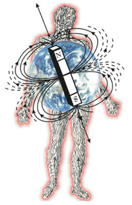
Magnetic Therapy in its natural state is
the dominant North Pole field of the Earth dominating over
the life processes of the body. With a full magnetic field
from the Earth the body goes through many actions to promote
good growth, strengthen tissue and fight disease and damage
from accidents or injury. However most of the Earth does
not have a full magnetic field. Scientists tell us that
as a normal process the Earth's poles reverse approximately
every 5,000 years. This could explain the loss of a lot
of dinosaur. As we move to this polarity switch, in about
2,000 years, the Earth's magnetic field is lessening. With
a decreased field the body is not always able to make all
the changes it should and it makes it unable to successfully
protect itself. An interesting note is that there are only
four places left on Earth with full magnetic fields. Two
habitable and two are not. The not are the North Pole and
the South Pole. The habitable are Sedona, Arizona and Lourdes,
France - both known for healthy living and healing. They
can be thought of as East and West Poles.
Magnetic Therapy works by affecting our blood.
Normally the blood operates in a North Pole orientation,
or under a North Pole effect. In this polarity the blood
is oxygenated and its process of distributing nutrients
and pulling wastes and toxins from injured tissue is made
most efficient. When the body is ill or injured the polarity
of the site is switched by the body to a South Pole orientation.
This creates faster, excited movement meant to draw blood
cells to the area for healing. The blood does not work
well in a South Pole orientation. Its movement does not
allow normal function and an acid state is developed, which
micro-organisms, viruses and malignance thrive in. *The
chart below shows the affect of North and South Pole applications
on the body.* Once the blood has been drawn to the area,
the body, with the help of the Earth's magnetic field is
supposed to change the polarity of the blood back to a
North Pole orientation so positive activity by the blood
may take place.
The problem is that with a reduced magnetic
field the body can not always make this necessary conversion
and the injury/illness area is left in a South Pole orientation
thwarting good cell growth with its acid effect and slowing
the healing process. Without blood removing wastes and
toxins from injury/illness sites they are left there to
fester and become unwanted bursas or arthritic tissue or
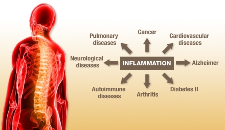
bad calcification. The increase in conditions like Arthritis,
Rheumatoid Arthritis, Fibromyalgia, ADD, ADHD and a multitude
of cancers and other Auto-Immune system diseases have increased
with the decrease of the magnetic field. Other problems
are slow healing in fracture sets and longer recovery periods
of disability after accidents. Another sign of this is
the great increase in repetitive motion injuries in every
form of business and sport. The answer in many cases is
Proper Polarity Magnetic Therapy.
By applying a structured North Pole magnetic
field, using high strength North Pole pad, we can convert
the polarity of the blood in the injury/illness area allowing
the blood to work as it should, pulling wastes and toxins
away to the kidneys and other cleansing organs, clearing
a path for good cell growth aided by the nutrients the
blood can now deliver. Simple and totally natural. Use
a Natural field to act as a catalyst to normal blood functions.
The body can now heal itself naturally. Of course if there
are broken bones or vertebrae out of place the magnets
will not do it alone, this is time for a good chiropractor.
Magnetic Therapy is only a piece of the health pie allowing
us to avoid in many cases unnatural medicines and sometimes
making surgical procedures unnecessary. Magnetic Therapy
works great on many conditions and studies are coming up
with more uses every day. Like the positive effects of
drinking North Pole magnetized water for those with conditions
like the Arthritis's, Fibromyalgia, Gout and ADHD to name
a few. People are having great success but that's not a
reason to avoid health professionals. After all without
good diagnosis what do we treat? Magnetic Therapy works
as well on Horses, Dogs and Cats as it does for people.
Apply a natural North Pole magnetic field.
Convert the polarity of the blood to the North Pole orientated
working polarity. Let the body heal itself. What is more
natural than that?
One of the most important series of experiments ever done
and yet still not widely known, were those carried out
by Albert Roy Davis in conjunction with Walter Rawls. These
experiments are FUNDAMENTAL to an understanding of magnetic
forces and are to this day not being used widely because
many have never heard of them.
They found that each pole of a magnet has SPECIFIC effects that are quite different
from those of a full magnet where both poles are applied simultaneously. These
polar effects are deemed "mono-polar"for one pole. The poles spin
in opposite directions and have opposite properties.
Specifically, North Pole energies cause mass to contract and condense, rotating
in a CCW direction, while South Pole energies cause mass to expand and dissipate,
rotating in a CW direction.
A new field of energy medicine is emerging. Magnetic energy
can be used as a non- nutritional source of energy to evoke
specific biological responses. Life is a matter of balance
between north and south pole magnetic polarities and not
just one or the other of the poles. The metabolic responses
of biological systems to specific magnetic energy polarity
and Gauss strength are consistently dependable. The body
is an electrical as well as a chemical unit. Magnetic fields
produce small electrical currents under the skin - sufficiently
powerful enough to cause biological effects such as pain
reduction, regeneration of cells and nerves, etc. Magnets
usually work better than they are predicted to - they are
the safest and finest therapy you can buy - nothing is
better in many cases. If you sense any discomfort while
wearing any magnetic device - take it off and the discomfort
will go away very quickly. You don't have to worry about
creating another conditions from an overdose
The beauty of the universe and the biological systems
within
it is its marvelous simplicity, unity of design and orderliness.
North magnetic energy can cause increased melatonin
production by stimulating the pineal gland at night.
Melatonin has been established as having the values of a
master control over hormones, sleep, immune
function, anti oxidants, infections, respiration, mineral
metabolism and all other
metabolic
energy systems. While our body is recovering its biological
energy loss, as we sleep, melatonin and growth hormone
are active participants in this recovery process
We hope this helps
provide a basic understanding how the Magnetic effect, especially
PEMFs, work in the body. In addition to these basic balancing
actions, magnetic fields also help with many other functions
and conditions, and new ones are discovered regularly.
Some of these other actions include:
•
reducing muscle tension
•
improving tissue healing
•
reducing pain
•
increasing energy
•
improving clotting factors
•
slowing the development of arthritis
•
stimulating the immune system
•
helping the body to detoxify
•
improving the uptake of nutrients
•
reducing blood pressure
•
helping nerve function
•
helping liver function
•
balancing the acupuncture meridians
•
improving sleep
•
making soft tissue more flexible
•
reducing arthritic changes
Selected Bibliography
Abadie, V., C. Gordon-Pomares, and J. Schirrer. “Analysis of the Olfactory
Capacity of Healthy Children Before Language Acquisition.” Journal of Developmental
and Behavioral Pediatrics 23.2 (August 2002): 203-207.
Addiction Science Research and Education Center, College
of Pharmacy, University of Texas. “Dopamine - A Sample
Neurotransmitter.” University of Texas. Academic,
2000. Available at http://www.utexas.edu/research/asrec/dopamine.html
(November 2004).
---. “Neurotransmitters Send Chemical "Messages".” University
of Texas. Academic, 2000. Available at http://www.utexas.edu/research/asrec/synapse.html
(November 2004).
---. “Other Neurostransmitters: Serotonin, Norepinephrine,
Acetylcholine, Glutamate and GABA (gamma-amino butyric
acid).” University of Texas. Academic, 2000. Available
at http://www.utexas.edu/research/asrec/synapse.html (November
2004).
Ahrens, B., B. Muller-Oerlinghausen, and M. Schou. “Excess
cardiovascular and suicide mortality of affective disorders
may be reduced by lithium prophylaxis.” Journal of
Affective Disorders 33 (1995): 67-75.
Akiskal, H.S., J. Downs, P. Jordan, S. Watson, D. Daugherty,
and D.B. Pruitt. “Affective disorders in referred
children and younger siblings of manic-depressives.” Archives
of General Psychiatry 42 (1985): 996-1003.
---, J.D. Maser, and P.J. Zeller. “Switching from
unipolar to bipolar II: an 11-year prospective study of
clinical and temperamental predictors in 559 patients.” Archives
of General Psychiatry 52 (1995): 114-123.
Anderson, C.A., and C.L. Hammen. “Psychosocial outcomes
of children of unipolar depressed, bipolar, medically ill,
and normal women: a longitudinal study.” Journal
of Consult. Clin. Psychol. 61 (1993): 448-454.
Anyanwu, E., G.F. Harding, A. Edson, and Department of
Vision Sciences, Aston University, Birmingham, UK. “The
involvement of serotonin (5-hydroxytryptamine) in photosensitive
epilepsy.” Journal of Basic Clinical Physiology and
Pharmacology 5.3-4 (1994): 179-206.
Appleton, R., M. Beirne, and B. Acomb. “Photosensitivity
in juvenile myoclonic epilepsy.” Seizure 9.2 (March
2000): 108-111.
Axel, Richard. “Representations of Olfactory Information
in the Brain.” Howard Hughes Medical Institute. Research
Foundation, October 2004. Available at http://www.hhmi.org/research/investigators/axel.html
(November 2004).
Bailey, C.H., and E.R. Kandel. “Structural changes
accompanying memory storage.” Annual Review of Physiology
55 (1993): 397-426.
Bateson, P.P.G. “The characteristics and context
of imprinting.” Biological Review 41 (1966): 177-220.
Beaulieu, C., and M. Colonnier. “Effect of the richness
of the environment on the cat visual cortex.” Journal
of Comparative Neurology 266 (1987): 478-494.
Becker, L.E., D.L. Armstrong, and F. Chan. “Dendritic
atrophy in children with Down syndrome.” Annual of
Neurology 20 (1986): 520-526.
Bennet, E., and M. Rosenzweig. “Behavioral and biochemical
methods to study brain responses to environment and experience.” In
Methods in Neurobiology. Vol. 2, 101-141. 1981.
Biederman, J., S.V. Faraone, K. Keenan, and M.T. Tsuang. “Evidence
of familial association between attention deficit disorder
and major affective disorders.” Archives of General
Psychiatry 48 (1991): 633-642.
---, J. Wozniak, and K Kiely. “CBCL clinical scales
discriminate prepubertal children with structured interview-derived
diagnosis of mania from those with ADHD.” Journal
of American Academic Childhood and Adolescent Psychiatry
34 (1995): 464-471.
Black, J.E., W.T. Greenough, B.J. Anderson, and K.R. Isaacs. “Environment
and the aging brain.” Canadian Journal of Psychology
41 (1987): 111-130.
---, M. Polinski, and W.T. Greenough. “Progressive
failure of cerebralangiogenesis supporting neural plasticity
in aging rats.” Neurobiological Aging 10 (1989):
353-358.
Bobula, B., A. Zahorodna, K. Tokarski, and G. Hess. “Use
of models for spontaneous paroxysmal activity in studies
of adaptive changes in serotonin receptors.” Postepy
higieny i medycyny doswiadczalnej 56.3 (2002): 377-383.
Borchardt, C.M., and G.A. Bernstein. “Comorbid disorders
in hospitalized bipolar adolescents compared with unipolar
depressed adolescents.” Childhood Psychiatry and
Human Development 26 (1995): 11-18.
Botteron, K.N., and B. Geller. “Pharmacologic treatment
of childhood and adolescent mania.” Psychiatric Clinics
of North America 4 (1995): 283-304.
---, M.W. Vannier, B. Geller, R.D. Todd, and B.C. Lee. “Preliminary
study of magnetic resonance imaging characteristics in
8- to 16-year-olds with mania.” Journal of American
Academic Childhood and Adolescent Psychiatry 34 (1995):
742-749.
Bottjer, S.W., and A.P. Arnold. “Developmental plasticity
in neural circuits for a learned behavior.” Annual
Review of Neuroscience 20 (1997): 459-481.
Brent, D.A., J.A. Perper, and C.E. Goldstein. “Risk
factors for adolescent suicide: a comparison of adolescent
suicide victims with suicidal inpatients.” Archives
of General Psychiatry 45 (1988): 581-588.
---, J.A. Perper, and G. Moritz. “Psychiatric risk
factors for adolescent suicide: a case-control study.” Journal
of Academic Childhood and Adolescent Psychiatry 32 (1993):
521-529.
---, J.P. Zelenak, O. Bukstein, and R.V. Brown. “Reliability
and validity of the structured interview for personality
disorders in adolescents.” Journal of Academic Childhood
and Adolescent Psychiatry 29 (1990): 349-354.
Brickner, D.D. “Educational synthesizer.” In
Hey, don't forget about me!, ed. A. Thomas. VA: Council
for Exceptional Children, 1976.
Brooks, P.H., and A.A. Baumeister. “A plea for consideration
of ecological validity in the experimental psychology of
mental retardation: A guest editorial.” American
Journal of Mental Deficiency 81 (1977): 407-416.
Browning, R.A., A.V. Wood, M.A. Merrill, J.W. Dailey,
and P.C. Jobe. “Enhancement of the anticonvulsant
effect of fluoxetine following blockade of 5-HT1A receptors.” European
Journal of Pharmacology 336.1 (October 1997): 1-6.
Bruell, S.J., and P.D. Coleman. “Dendritic growth
in the aged human brain and failure of growth in senile
dementia.” Science 206 (1979): 854-856.
--- and P.D. Coleman. “Regulation of dendritic extent
in developing and aging brain.” In Synaptic Plasticity,
ed. C.W. Cotman, 311-333. New York: Raven, 1985.
Brunet, and Lézine. Echelle de Development Psychomoteur
de la Première Enfance, rev. ed. Paris: Les Editions
du Centre de Psychologies Appliquées, 1998.
Brunjes, P.C., and L.L. Frazier. “Maturation and
plasticity in the olfactory system of vertebrates.” Brain
Research Review 11 (1986): 145.
Calvin, W.H. The Cerebral Code. Cambridge, MA: MIT Press,
1996.
Carlson, G.A., and J.H. Kashani. “Manic symptoms
in a non-referred adolescent population.” Journal
of Affective Disorders 15 (1988): 219-226.
Cattell, P. Infant Intelligence Scale. New York: Psychological
Corp., 1940.
Chaouloff, F., and . Serotonin regulates stress. Bordeaux,
France: INSERM U47 1, Institut Megendie, .
Childs, B., and C.R. Scriver. “Age at onset and
causes of disease.” Perspectives in Biology and Medicine
29 (1986): 437-460.
Chugani, D.C., and Departments of Pediatrics and Radiology,
Children's Hospital of Michigan, Detroit Medical Center,
Wayne State University School of Medicine, Detroit, Michigan. “Serotonin
in autism and pediatric epilepsies.” Mental Retardation
and Developmental Disabilities Research and Development
10.2 (2004): 112-116.
Clinckers, R., I. Smolders, A. Meurs, G. Ebinger, and
Y. Michotte. “Anticonvulsant action of hippocampal
dopamine and serotonin is independently mediated by D and
5-HT receptors.” Journal of Neurochemistry 89.4 (May
2004): 834-843.
Compan, V., M. Zhou, R. Grailhe, R. A. Gazzara, R. Martin,
and J. Gingrich. “Attenuated response to stress and
novelty and hypersensitivity to seizures in 5-HT4 receptor
knock-out mice.” Journal of Neuroscience 24.2 (January
2004): 412-419.
Cooper, T.B., P.E. Bergner, and G.M. Simpson. “The
24-hour serum lithium level as a prognosticator of dosage
requirements.” American Journal of Psychiatry 130
(1973): 601-603.
Coryell, W., J. Endicott, J.D. Maser, M.B. Keller, A.C.
Leon, and H.S. Akiskal. “Long- term stability of
polarity distinctions in the affective disorders.” American
Journal of Psychiatry 152 (1995): 385-390.
---, M. Keller, J. Endicott, N. Andreasen, P. Clayton,
and R. Hirschfeld. “Bipolar II illness: course and
outcome over a five-year period.” Psychol. Med. 19
(1989): 129-141.
---, W. Scheftner, M.B. Keller, J. Endicott, J. Maser,
and G.L. Klerman. “The enduring psychosocial consequences
of mania and depression.” American Journal of Psychiatry
150 (1993): 720-727.
Crowell, M. D. “The role of serotonin in the pathophysiology
of irritable bowel syndrome.” American Journal of
Managed Care 7.Suppl 8 (July 2001): 252-260.
Davis, J. M., N. L. Alderson, and R. S. Welsh. “Serotonin
and central nervous system fatigue: nutritional considerations.” American
Journal of Clinical Nutrition 72.Suppl 2 (August 2000):
573-578.
Decina, P., C.J. Kestenbaum, and S. Farber. “Clinical
and psychological assessment of children of bipolar probands.” American
Journal of Psychiatry 140 (1983): 548-553.
Delgado, P.L., and F.A. Moreno. “Hallucinogens,
serotonin and obsessive-compulsive disorder.” Journal
of Psychoactive Drugs 30.4 (1998): 359-366.
Di Matteo, V., A. De Blasi, C. Di Giulio, and E. Esposito. “Role
of 5-HT(2C) receptors in the control of central dopamine
function.” Trends in Pharmacological Science 22.5
(May 2001): 229-232.
Dominquez, H.D., M.F. Lopez, and J.C. Molina. “Interactions
between perinatal and neonatal associative learning defined
by contiguous olfactory and tactile stimulation.” Neurobiological
Learning Memory 71.3 (1999): 272-288.
DuBose, R.F., and M.B. Langley. The developmental activities
screening inventory. Boston: Teaching Resources, 1977.
Dugovic, C. “Role of serotonin in sleep mechanisms.” Review
of Neurology (Paris) 157.11 (November 2001): 16-19.
Fetner, H.H., and B. Geller. “Lithium and tricyclic
antidepressants.” Psychiatric Clinics of North America
15 (1992): 223-224.
Fewell, R.R., and University of Miami School of Medicine. “Play
assessment scale.” 1992.
---P.F. Vadasy. “Supports from religious organizations
and personal beliefs.” In Families of handicapped
children: Needs and supports across the life span, ed.
R.R. Fewell, 297-316. Austin, TX: PRO-ED, 1986.
---D. Weatherford. “The measurement of family functioning.” In
Evaluating early intervention programs for severely handicapped
children and their families, ed. L. Bickman, 263-307. Austin,
TX: PRO-ED, 1986.
Folio, M.R., and R.F. DuBose. “Peabody developmental
motor scales (Monograph No. 25).” , George Peabody
College, IMRID Behavioral Research Institute, 1974.
Fraiberg, S.M. The magic years. New York: Charles Scribner's
Sons, 1959.
Gage, Fred H. “BRAIN, REPAIR YOURSELF. How do you
fix a broken brain? The answers may literally lie within
our heads. The same approaches might also boost the power
of an already healthy brain.” Scientific American
(September 2003).
Gesell, A., and C.S. Amatruda. Developmental diagnosis,
2nd ed. New York: Paul B. Hoeber, 1947.
Gilliam, F. G., J. Santos, V. Vahle, J. Carter, K. Brown,
and H. Hecimovic. “Depression in epilepsy: ignoring
clinical expression of neuronal network dysfunction?” Epilepsia
45.Suppl 2 (2004): 28-33.
Gordon-Pomares, C. L’Eveil Sensoriel de Mon Enfant.
Paris: Hachette, 1995.
---. Manuel Pratique du Jouet. Paris: Hachette, 1998.
---. Papa, Maman, L’Ecole et Moi. Paris: Hachette,
1997.
Gray, J. A., and B. L. Roth. “Paradoxical trafficking
and regulation of 5-HT(2A) receptors by agonists and antagonists.” Brain
Research Bulletin 56.5 (15 November 2001): 441-451.
Grimaldi, B., A. Bonnin, M. P. Fillion, N. Prudhomme,
and G. Fillion. “5-Hydroxytryptamine-moduline: a
novel endogenous peptide involved in the control of anxiety.” Neuroscience
93.4 (1999): 1223-1225.
--- and G. Fillion. “5-HT-moduline controls serotonergic
activity: implication in neuroimmune reciprocal regulation
mechanisms.” Progress in Neurobiology 60.1 (January
2000): 1-12.
Haeussermann, E. Developmental potential of preschool
children. New York: Grune & Stratton, 1958.
Harvey, J.A. “Serotonin & Memory/Review: Role
of the Serotonin 5-HT2A Receptor in Learning.” ,
Dept of Pharmacology and Physiology, Laboratory of Behavioral
Neurobiology, Drexel University College of Medicine, Philadelphia,
.
Heisler, L. K., M. A. Cowley, T. Kishi, L. H. Tecott,
W. Fan, and M. J. Loe. “Central serotonin and melanocortin
pathways regulating energy homeostasis.” Annals of
the New York Academy of Sciences 994 (June 2003): 169-174.
Holloway, Marguerite. “THE MUTABLE BRAIN. Score
one for believers in the adage "Use it or lose it." Targeted
mental and physical exercises seem to improve the brain
in unexpected ways .” Scientific American (September
2003).
Holsboer, F. “Stress, hypercorticolism and corticosteroid
receptors in depression: implications for therapy.” Journal
of Affective Disorders 62 (2001): 77-91.
Horvitz, J.C. “Dopamine.” http://www.columbia.edu.
Academic, 2000. Available at http://www.columbia.edu/~jh299/DA.html#1989
(November 2004).
---. “Mesolimbic and nigrostriatal dopamine responses
to salient non-reward events.” Neuroscience 96.4
(March 2000): 651-656.
--- and Y.S. Eyny. “Dopamine D2 receptor blockade
reduces response likelihood but does not affect latency
to emit a learned sensory-motor response: Implications
for parkinson's disease.” Behavioral Neuroscience
114.5 (2000): 934-939.
---, W.B. Richardson, and A. Ettenberg. “Dopamine
receptor blockade and thirst produce differential effects
on drinking behavior.” Pharmacology Biochemistry
and Behavior 45 (1993): 725-728.
---, T. Sterwart, and B.L. Jacobs. “Burst activity
of ventral tegmental dopamine neurons is elicited by sensory
stimuli.” Brain Research 759 (1997): 251-258.
Horwitz, J.C., T. Sterwart, B.L. Jacobs, and . “Burst
activity of ventral tegmental dopamine neurons is elicited
by sensory stimuli.” Brain Research 759 (1997): 251-258.
Johnson, B.A., S. Ho, Z. Xu, J.S. Yihan, S. Yip, and E.E.
Hingco. “Functional mapping of the rat olfactory
bulb using diverse odorants reveals modular responses to
functional groups and hydrocarbon structural features.” Journal
of Comparative Neurology 449 (2002): 180-194.
--- and M. Leon. “Modular glomerular representations
of odorants in the rat olfactory bulb and the effects of
stimulus concentration.” Journal of Comparative Neurology
422 (2000): 496-509.
--- and M. Leon. “Odorant molecular length: One
aspect of the olfactory code.” Journal of Comparative
Neurology 426 (2000): 330-338.
---, C.C. Woo, E.E. Hingco, K.L. Pham, and M. Leon. “Multidimensional
chemotopic responses to n-aliphatic acid odorants in the
rat olfactory bulb.” Journal of Comparative Neurology
409 (1999): 529-548.
---, C.C. Woo, and M. Leon. “Spatial coding of odorant
features in the glomerular layer of the rat olfactory bulb.” Journal
of Comparative Neurology 393 (1998): 457-471.
Kolb, B., M. Forgie, R. Gibb, G. Gorny, and S. Rowntree. “Age,
experience and the changing brain.” Neuroscience
and Behavioral Review 22 (1998): 143-159.
---, R. Gibb, and G. Gorny. “Cortical plasticity
and the development of behavior after frontal cortical
injury.” Developmental Neuropsychology 18 (2000):
423-444.
---, R. Gibb, and G. Gorny. “Experience-dependent
changes in dendritic arbor and spine density in neocortex
vary with age and sex..” Neurobiolgy of Learning
and Memory 79 (2003): 1-10.
--- and I.Q. Wishaw. “Brain Plasticity and Behavior.” Annual
Review of Psychology 49 (1998): 43-64.
LaHoste, G.J., and . “Dopamine D4 receptor gene
polymorphism is associated with attention-deficit hyperactivity
disorder.” Molecular Psychiatry 1.2 (May 1996): 121-124.
Langdon, J., and H. Down. “OBSERVATIONS ON AN ETHNIC
CLASSIFICATION OF IDIOTS.” Clinical lectures and
reports by the medical and surgical staff of the London
Hospital 3 (1866): 259-62.
Lawrence, T.M. “Precision teaching in perspective:
An interview with O. R. Lindsley.” Teaching Exceptional
Children 3.3 (1971): 114-119.
Leon, M., R. Coopersmith, F.B. Weihmuller, C.L. Kirstein,
and J.F. Marshall. “Extracellular dopamine increases
in the neonatal olfactory bulb during odor preference training.” Brain
Research 564.1 (1991): 149-153.
Linster, C., B.A. Johnson, A. Morse, E. Yue, and M. Leon. “Spontaneous
vs. reinforced olfactory discriminations.” Journal
of Neuroscience 22 (2002): 6842-6845.
---, B.A. Johnson, A. Morse, E. Yue, Z. Xu, and Y. and
M. Choi. “Perceptual correlates of neural representations
evoked by odorant enantiomers.” Journal of Neuroscience
21 (2001): 9837-9843.
Liu, Diorio, Tannenbaum, Caldji, Francis, and Freedman. “Maternal
Care, Hippocampal Glucocorticoid Receptors, and Typothalamic-Pituitary-Adrenal
Responses to Stress.” Science 277 (1997): 1659-1662.
Mann, J., A. McBride, and R. Brown. “Relationship
between central and peripheral serotonin indexes in depressed
and suicidal psychiatric inpatients.” Archives of
General Psychiatry 49 (1992): 442-446.
Matsumoto, M., and M. Yoshioka. “Possible involvement
of serotonin receptors in anxiety disorders.” Nippon
Yakurigatu Zasshi 2000.115 (1): 39-44.
McEwen, B.S. “Protective and damaging effects of
stress mediators.” New England Journal of Medicine
338.3 (1998): 171-179.
McLean, J.H., and A. Darby-King. “Receptor involvement
in conditioned olfactory learning in the neonate rat pup.” Behavioral
Neuroscience 110 (1996): 1426-1434.
---, S. Darby-King, R.M. Sullivan, and S.R. King. “Serotonergic
influence on olfactory learning in the neonate rat.” Behavioral
Neural Biology 60.2 (1993): 152-162.
Meneses, A. “Endogenous serotonin (5-hydroxytryptamine
[5-HT]) modulates plasticity processes, including learning
and memory.” , Department of Pharmacobiology, CINVESTAV-IPN,
Mexico, .
--- and Departamento de Farmacologia y Toxicologia, CINVESTAV-IPN,
Mexico D.F., Mexico. “Physiological, pathophysiological
and therapeutic roles of 5-HT systems in learning and memory.” Review
of Neuroscience 9.4 (1998): 275-289.
--- and Departamento de Farmacologia y Toxicologia, CINVESTAV-IPN,
Mexico D.F., Mexico. “5-HT system and cognition.” Neuroscience
and Biobehavioral Reviews. 23.8 (1999): 1111-1125.
Merlet, I., K. Ostrowsky, N. Costes, P. Ryvlin, J. Isnard,
and F. Lavenne. “5-HT1A receptor binding and intracerebral
activity in temporal lobe epilepsy: an PET study.” Brain
127.4 (April 2004): 900-913.
---, P. Ryvlin, N. Costes, D. Dufournel, J. Isnard, and
K. Ostrowsky. “Statistical parametric mapping of
5-HT1A receptor binding in temporal lobe epilepsy with
hippocampal ictal onset on intracranial EEG.” Neuroimage
22.2 (June 2004): 886-896.
Mitchell, J.B., and A. Gratton. “Mesolimbic dopamine
release elicited by activation of the accessory olfactory
system: a high-speed chronoamperometric study.” Neuroscience
Letter 140.1 (1992): 81-84.
Mombaerts, P., F. Wang, C. Dulac, R. Vassar, S.K. Chao,
A. Nemes, and Department of Biochemistry and Molecular
Biophysics, Howard Hughes Medical Institute, College of
Physicians and Surgeons, Columbia University, New York. “The
molecular biology of olfactory perception.” Cold
Spring Harb Symp. Quant. Biol. 61 (1996): 135-145.
Murphy, C., and S. Jinich. “Olfactory dysfunction
in Down syndrome.” Neurobiological Aging 17.4 (1996):
631-637.
Nadi, N.S., R. Head, M. Grillo, J. Hempstead, N. Grannot-Reisfeld,
and F.L. Margolis. “Chemical deafferentation of the
olfactory bulb: plasticity of the levels of?tyrosine hydroxylase,
dopamine and norepinephrine..” Brain Research.213
(June 1981), 365-377.
Naughton, M., J.B. Mulrooney, and B.E. Leonard. “A
review of the role of serotonin receptors in psychiatric
disorders.” Human Psychopharmacology 15.6 (2000):
397-415.
Philpot, B.D., D. Men, R. McCarty, and P.C. Brunjes. “Activity-dependent
regulation of dopamine content in the olfactory bulbs of
naris-occluded rats.” Journal of Neuroscience 85.3
(1998): 969-977.
Pitts, S.M., and J.C. Horvitz. “Similar effects
of D1/D2 receptor blockade on feeding and locomotor behavior.” Pharmacology
Biochemistry and Behavior 65 (2000): 433-438.
Rangel, S., and M. Leon. “Early odor preference
training increases olfactory bulb norepinephrine.” Journal
of Developmental Brain Research 85.2 (1995): 187-191.
Robbins, N. “The teaching of a manual sign as a
diagnostic tool with deaf-blind children.” In Fourth
International Conference on Deaf-Blind Children. Waterton,
MA: Perkins School for the Blind, 1971.
Rosenzweig, M. “Experience, memory and the brain.” Journal
of American Psychology 39.4 (1984): 365-376.
---, E. Bennet, and J. Flood. “Pharmocological modulation
of formation of long-term memory..” In Brain and
Behavior. Vol. 17, 101-111. 1981.
Royet, J.P., F. Jourdan, H Ploye, and C Souchier. “Morphometric
modifications associated with early sensory experience
in the rat olfactory bulb: II. Stereological study of the
population of olfactory glomeruli.” Journal of Comparative
Neurology 289.4 (1989): 594-609.
Saito, H., M. Matsumoto, H. Togashi, and M. Yoshioka. “Functional
interaction between serotonin and other neuronal systems:
focus on in vivo microdialysis studies.” Japanese
Journal of Pharmacology 70.3 (March 1996): 203-205.
Sapolsky, Robert. “TAMING STRESS. An emerging understanding
of the brain's stress pathways points toward treatments
for anxiety and depression beyond Valium and Prozac .” Scientific
American (September 2003).
Schaal, B., H. Montagner, E. Hertling, D. Bolzoni, A.
Moyse, and R. Quichon. “Les stimulations olfactives
dans les relations entre l’enfant et la mère.” Nutrition
and Development 20.3B (1980): 843-858.
Sevcik, J., P. Petrovicky, V. Ruzicka, J. Slansky, and
K. Masek. “Structures and pathways of the central
nervous system are potentially involved in the serotonergic
modulation of gastrointestinal activity.” Methods
and findings in experimental and clinical pharmacology.
24.10 (2002): 669-673.
Silberstein, S. D. “Serotonin (5-HT) and migraine.” Headache
34.7 (1994): 408-417.
Southwick, S. M., S. Paige, C. A. Morgan, J. D. Bremner,
J. H. Krystal, and D. S. Charney. “Neurotransmitter
alterations in PTSD: catecholamines and serotonin.” Seminars
in clinical neuropsychiatry 4.4 (October 1999): 242-248.
Steiger, H., M. Isra‘l, N. Koerner, N. Kin, J. Paris,
and S.N. Young. “Bulimia nervosa (BN) is reported
to co-occur with childhood abuse and alterations in central
serotonin (5-hydroxytryptamine [5-HT]) and cortisol mechanisms.” Archives
of General Psychiatry 58 (2001): 837-843.
Steinkruger, M. “Photosensitive epilepsy.” Journal
of Neurosurgical Nursing 17.6 (1985): 355-361.
Steriade, M. “Slow-wave sleep: serotonin, neuronal
plasticity, and seizures.” Archives Italiennes de
Biologie 142.2 (July 2004): 359-367.
Stutsman, R. Merrill Palmer scale of mental tests. Chicago:
Stoelting, 1948.
Szatmari, Peter. “The Epidemiology of Attention-Deficit
Hyperactivity Disorder.” In Child and Adolescent
Pcychiatric Clinics of North America, ed. W.B. Saunders
and G. Weiss. Vol. 1. 1992.
Terron, J.A. “Is the 5-HT(7) receptor involved in
the pathogenesis and prophylactic treatment of migraine?” European
Journal of Pharmacology 439.1-3 (29 March 2002): 1-11.
Theodore, W.H. “Does Serotonin Play a Role in Epilepsy?” Epilepsy
Current 3.5 (September 2003): 173-177.
Trottier, S., B. Evrard, J. P. Vignal, J. M. Scarabin,
and P. Chauvel. “The serotonergic innervation of
the cerebral cortex in man and its changes in focal cortical
dysplasia.” Epilepsy Research 25.2 (October 1996):
79-106.
Van Esch, H., M. Syrrou, and L. Lagae. “Refractory
photosensitive epilepsy associated with a complex rearrangement
of chromosome 2.” Neuropediatrics 33.6 (December
2002): 320-323.
Van Heeringen, C. “Suicide, Serotonin and the Brain.” Crisis
22.2 (2001): 66-70.
Van Hooft, J. A., and H. P. Vijverberg. “5-HT(3)
receptors and neurotransmitter release in the CNS: a nerve
ending story?” Trends in Neuroscience 23.12 (2000):
605-610.
Villalon, C.M., D. Centurion, L.F. Valdivia, P. De Vries,
and P.R. Saxena. “An introduction to migraine: from
ancient treatment to functional pharmacology and antimigraine
therapy.” Proceedings of the Western Pharmacology
Society 45 (2002): 199-210.
Welsh, J.P., B. Chang, M.E. Menaker, S.A. Aicher, and
. “Removal of the inferior olive abolishes myoclonic
seizures associated with a loss of olivary serotonin.” Neuroscience
82.3 (1998): 879-897.
---, D.G. Placantonakis, S.I. Warsetsky, R.G. Marquez,
L. Bernstein, and S.A. Aicher. “Serotonin hypothesis
of myoclonus from the perspective of neuronal rhythmicity.” Advances
in Neurology 89 (2002): 307-329.
Will, B., and M. Rosenzweig. “Effects of differential
environments on recovery from neonatal brain lesions, measured
by problem solving scores.” Physiology and Behavior
16 (1976): 603-611.
Wood, B.L., S. Haque, A. Weinstock, B.D. Miller, and Division
of Child and Adolescent Psychiatry, State University of
New York at Buffalo, Buffalo, NY. “Pediatric stress-related
seizures: conceptualization, evaluation, and treatment
of nonepileptic seizures in children and adolescents.” Current
Opinions in Pediatrics 16.5 (2004): 523-531.
Yan, X.X., J. Najbauer, C.C. Woo, K. Dashtipour, C.E.
Ribak, and M. Leon. “Expression of active caspase-3
in mitotic and postmitotic forebrain cells of rat.” Journal
of Comparative Neurology 433 (2001): 4-22