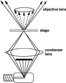
Dark Field Microscope
What is Blood and What Does it Do
Two types of blood vessels carry blood throughout our
bodies: The arteries carry oxygenated blood (blood that
has received oxygen from the lungs) from the heart to the
rest of the body.
The blood then travels through the veins back to the
heart and lungs, where it receives more oxygen. As the
heart beats, you can feel blood traveling through the body
at your pulse points - like the neck and the wrist - where
large, blood-filled arteries run close to the surface of
the skin.
The blood that flows through this network
of veins and arteries is called whole blood. Whole blood
contains three types of blood cells:
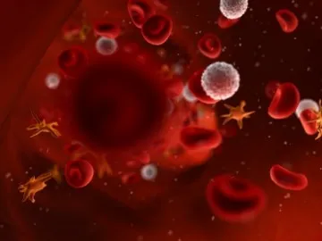
Red Blood Cells
White Blood Cells
Platelets
|
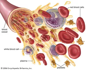 |
These blood cells are mostly manufactured
in the bone marrow (the soft tissue inside our bones),
especially in the bone marrow of the vertebrae (the bones
that make up the spine), ribs, pelvis, skull, and sternum
(breastbone). These cells travel through the circulatory
system suspended in a yellowish fluid called plasma (pronounced:
plaz-muh). Plasma is 90% water and contains nutrients,
proteins, hormones, and waste products. Whole blood is
a mixture of blood cells and plasma.
| Red Blood Cells |
 |
Red blood cells (RBCs, and also called erythrocytes,
pronounced: ih-rith-ruh-sytes) are shaped like slightly
indented, flattened disks. Red blood cells contain an iron-rich
protein called hemoglobin (pronounced: hee-muh-glow-bun).
Blood gets its bright red color when the hemoglobin in
RBCs picks up oxygen in the lungs. As the blood travels
through the body, the hemoglobin releases oxygen to the
tissues. The body contains more RBCs than any other type
of cell, and each has a life span of about 4 months. Each
day, the body produces new RBCs to replace those that die
or are lost from the body.
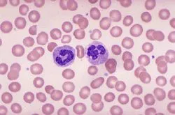
| White Blood Cells |
 |
White blood cells (WBCs, and also called leukocytes, pronounced:
loo-kuh-sytes) are a key part of the body's system for
defending itself against infection. They can move in and
out of the bloodstream to reach affected tissues. The blood
contains far fewer white blood cells than red cells, although
the body can increase production of WBCs to fight infection.
There are several types of white blood cells, and their
life spans vary from a few days to months. New cells are
constantly being formed in the bone marrow.
Several different parts of blood are involved in fighting
infection. White blood cells called granulocytes (pronounced:
gran-yuh-low-sytes) and lymphocytes (pronounced: lim-fuh-sytes)
travel along the walls of blood vessels. They fight germs
such as bacteria and viruses and may also attempt to destroy
cells that have become infected or have changed into cancer
cells.
Certain types of WBCs produce antibodies, special proteins
that recognize foreign materials and help the body destroy
or neutralize them. Someone with an infection will often
have a higher white cell count than when he or she is well
because more WBCs are being produced or are entering the
bloodstream to battle the infection. After the body has
been challenged by some infections, lymphocytes "remember" how
to make the specific antibodies that will quickly attack
the same germ if it enters the body again.
| Platelets |
 |
Platelets (also called thrombocytes, pronounced:
throm-buh-sytes) are tiny oval-shaped cells made in the
bone marrow. They help in the clotting process. When a
blood vessel breaks, platelets gather in the area and help
seal off the leak. Platelets survive only about 9 days
in the bloodstream and are constantly being replaced by
new cells.
Blood also contains important proteins called
clotting factors, which are critical to the clotting process.
Although platelets alone can plug small blood vessel leaks
and temporarily stop or slow bleeding, the action of clotting
factors is needed to produce a strong, stable clot.
Platelets and clotting factors work together to form solid
lumps to seal leaks, wounds, cuts, and scratches and to
prevent bleeding inside and on the surfaces of our bodies.
The process of clotting is like a puzzle with interlocking
parts. When the last part is in place, the clot happens
- but if only one piece is missing, the final pieces can't
come together.
When large blood vessels are severed (or cut), the body
may not be able to repair itself through clotting alone.
In these cases, dressings or stitches are used to help
control bleeding.
In addition to the cells and clotting factors, blood contains
other important substances, such as nutrients from the
food that has been processed by the digestive system. Blood
also carries hormones released by the endocrine glands
and carries them to the body parts that need them.
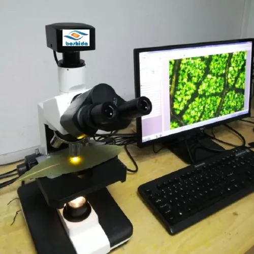
Now
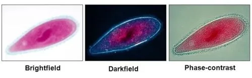
you can View your Blood Alive
Dark Field Microscope with HD camera
The advantage of darkfield illumination is
that you can see details that are normally not resolved
by the microscopes objective. You can't normaly see the
actual detail but because it reflects light you can . A
nice analogy is that of dust in a room. In a well lit room
you do not see the very small dust particles. However,
if the lights go out, a beam of light from an acute angle
makes these same particles visible. Besides the optical
advantages darkfield illumination is very beautiful and
gives an almost science fiction like image.
Darkfield blood analysis uses
a high-definition microscope to analyze the blood.
This method is very useful for the early detection of
serious health conditions.
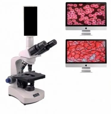
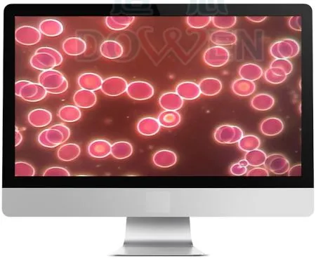
As the blood is being analyzed under the microscope the
images are being passed to a monitor screen where You
or a practitioner can analyze and
discuss the patient's blood in its living state.
It is a way to look at one drop of
your living blood from a simple prick of your finger.
That drop is placed on a slide, enlarged and projected
onto a monitor. The invisible comes alive as you instantly
enter the world of your living blood.
The
same microscopic equipment utilized in the Live
Blood Analysis screening test is also used
in the Dry Blood Test.
Both tests use droplets of blood expressed
from the tip of the finger. |
Traditional blood tests
use a light source so hot that it kills the blood, and
a stain that is looking only for a particular microbe
- it is an autopsy of your blood.
Blood Under a Microscope
Dark field is used for Initial examination
of suspensions of cells such as yeast, parasites,bacteria,
small protists, or cell and tissue fractions including
cheek epithelial cells, chloroplasts, mitochondria, even
blood cells Pics
Of Course,The FDA does not approve of dark field
microscopic blood analysis, therefore many doctor's
hands are tied. Viewing a fresh, natural blood sample
(a sample not altered with any stains, etc., needed
for normal microscopic exams), under the technology
of a dark field microscope, will reveal conditions
of your blood not normally even considered during
the diagnosis of a normal blood test performed in
doctor's office or a lab.
However, an increasing
number of health professionals have found that
the use of this technique allows inspection
of cellular dynamics which as noted above normally
escape analysis or diagnosis using orthodox
medical tests.
|
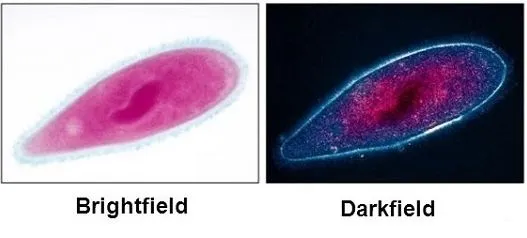
Darkfield microscopes employ a special contrast
enhancing technique known as dark field illumination to
produce beautiful images of normally difficult-to-observe
biological specimens. Similar to the phenomenon of being
able to see stars at night but not during the day, darkfield
illumination is most often used with samples that are not
easily imaged against a light background, and results in
samples that appear bright against a dark background
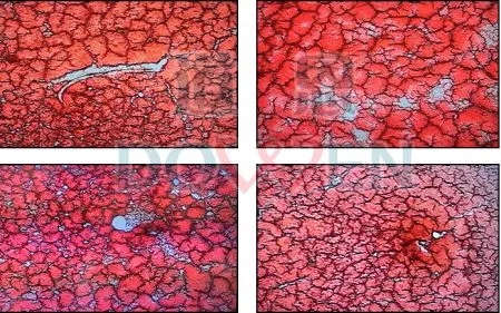
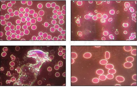
How
to Interpret what you see
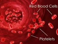
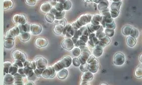
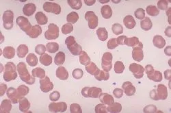
 Healthy Blood Cells
 |
UnHealthy Blood Cells
|
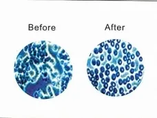
| Do you see how far apart
the blood cells are from each other? As a result, your blood
can move freely throughout your entire body, and
get into all your small capillaries, providing energy
to your whole body. During deep sleep, proper blood
flow and hydration is important. When your blood looks
like this, your sleep is also really energizing and
you need less of it! |
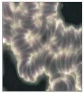
When your blood
is clumped together, it no longer can get to
all the little capillaries in your body to give you
the life giving oxygen you need. It no longer can
give every cell of your body the energizing and rejuvenating
effects. This is the major reason why some people
feel horrible when they wake up, and why they need
to sleep longer. It's also why you tend to wake up
feeling dehydrated. |
In darkfield microscopy, one is therefore able to observe "live
blood." Unlike the techniques of electron microscopy,
no fixative is used so the picture is one of mobility rather
than fixity. With stains and fixatives, the picture reveals
a moment in time rather than a continuum.
What
Makes Up Healthy Blood?
What one sees in the mobile situation are the usual red
blood cells, white blood cells, plasma—and what is
floating in the plasma. Microbial activity, undigested
food, fungi, and crystals are all apparent as is the capacity
of the red blood cells to circulate and the white blood
cells to devour morbid matter.
Darkfield Microscopy
or Live Blood Analysis
(Live Blood Examination in the Darkfield according to Prof. Dr. G. Enderlein)
Darkfield Microscopy or Live Blood Analysis is a way of studying live whole
blood cells under a specially adapted microscope that projects the dynamic
image onto a video screen. This allows you to view your inner terrain. Digestive,
eliminative and immune functions can be assessed as well as the presence of bacteria
and other micro-organisms.
The darkfield microscopic examination of the freshly taken
live blood is one of the most important examinations of
the holistic medicine applied at the Centre. It enables
us to view the inner terrain (milieu) and to examine the
functions of the red blood cells. It also shows the evolutionary
stages of the smallest proteins (endobionts) which are
found in every human body. We are also able to see any
developed structures such as bacteria, virus and fungus.
The darkfield examination shows the state of the blood
cells, endobionts and the plasma in a functional and structural
way, making bacterial processes and fungal pre-stages in
the blood clearly visible.
| Most of the time, blood functions
normally, but sometimes, blood disorders or diseases
can cause problems. Diseases of the blood that
commonly affect people can involve any or all of
the three types of blood cells (red blood cells,
white blood cells, or platelets) or the proteins
and chemicals in the plasma that are responsible
for clotting.More |
The darkfield
examination is most suitable for the evaluation of
chronic diseases; for children who are prone to infections;
for recurrent bacterial problems; for candida and other
fungal problems and also to answer questions concerning
chronic problems of toxicity (e.g. amalgam disturbances).
Some Interesting
Uses for Darkfield Microscopes you can Experiment With
Water, Consciousness & Intent:
Dr. Masaru Emoto
|
Microscope Video of a Drop of Pond Water
|
| He freezes droplets of water
and then examines them under a dark field microscope
that has photographic capabilities.More |
Suspensions of cells and
samples of pond water look spectacular in dark field.
While specimens may look washed out and lack detail
in bright field, protists, metazoans, cell suspensions,
algae, and other microscopic organisms are clearly
distinguished and their details show up well. At 100x
you can readily see bacteria, even distinguish some
structure (rods, curved rods, spirals, or cocci) and
movement. Non-motile bacteria look like vibrating bright
dots against a dark background. Motile bacteria can
be seen moving in a definite direction, sometimes remarkably
fast. In pond water samples you may find Spirillum
volutans, a very large (up to 0.5 mm) motile spiral
bacterium. |
Using a very fine needle, a drop of blood is taken from
the finger and directly placed on a glass slide. Without
fixation or colouring, the blood is examined right after
taking it through a special darkfield microscope with up
to 100x enlargement. You can follow the process via video
or Computer screen. The blood can be examined again several
hours after taking the sample. This procedure informs us
about the speed of degeneration of the cells (shows cell
resilience, the immune system and the degenerative tendency).How
to Interpret what you see
This examination was developed and described by Prof.
Dr. G. Enderlein. With this method he proved that co-relations
exist between blood parasites, symbionts, bacteria
and fungi. The main proven fact is that chronic diseases
are created by increasing
sickness tendencies of the endobionts and
that bacteria, viruses and fungi developed in the human
body, or are changed to pathogenic agents of diseases depending
upon the inner terrain (determined by acid-base balance,
protein content and level of trace elements). The existence
of pre-stages which are not yet able to make one ill but
that can endanger an illness can also be found in the darkfield
examination. Therefore it is also an important preventative
tool.
The medical establishment has generally not been
keen to support the concept of viewing live blood
as a method of diagnosing and determining the health
of a patient. In many instances, they completely
omit and reject darkfield microscopy as a diagnostic
instrument. This same medical establishment has also
been resistant to natural health including the use
of herbs and natural supplements as an alternative
to the pushing of powerful pharmaceutical medical
drugs. It is acknowledged by this author that there
are certainly quack doctors out there that will use
whatever instrument they can to make a fast dollar. |
|
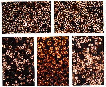
Many cancer patients have poor oxygenation
of their blood. Low hemoglobin, clumped red blood cells
(rouleau), infections, and toxicity can affect oxygenation,
vitality, and health. The picture to the left vividly
demonstrates how impossible it is for the red blood
cells to circulate and transport oxygen. From a health
perspective, this condition is an accident waiting
to happen. |
Dr Young is a world reknowned scientist and microbiologist
who studies live blood cells under the microscope.
He has found that excess acidity in the body causes
health problems and symptoms. Our blood in order to
remain healthy has to remain at a pH of 7.36 |
Laser
Blood Cleanser
Every once in a while a technology is introduced
that transforms the landscape of health care.
Low Level Laser Therapy is a perfect example
of such a development. It is well accepted,
painless and, bottom line, it gets results.
Laser Blood Cleanse Dark Field Results
More
Prices |
 |
|
 Blood
under darkfield. Microscopic examination which is moderately
to strongly infested
(source: Blood examination in darkfield, Semmelweis Verlag 1993 ISBN 3-925524-01-0) (Blood examination according to Prof. Dr. G. Enderlein) Darkfield Microscopy
is an important holistic diagnostic instrument used in practice.
It gives information about the terrain, e.g. hyperacidity, lack of energy
and dynamics and the functionality of blood cells.
Prof. Dr. G. Enderlein (1872-1968) observed in darkfield microscopic
examination of blood the tiniest moving beings , which seem to be primitive
forms of either bacteria or fungi.
These so-called Endobiont change the hydrogen-ion con centration (pH)
of the blood. Also Intoxication such as with heavy metal (e.g. mercury)
can be made visible.
For the examination we need only a drop of blood from the fingertip
or the ear. Observation of the blood sample in the darkfield microscope
gives information within 15 min. after hours or even days we find further
information regarding the condition of the immune system, cellular resistance
and the disposition towards the growing or regeneration of possible tumors.
Cancer, for example, cannot be seen but the weakness of the immune system
or the degeneration of the blood cells can be made visible which is invaluable
in early treatment.
More Pics |
Cleanse
The Blood
The Bob Beck Protocol Simply Put
The
Blood zapper emits pulsed micro-amps causing
the blood and tissue cell membranes to
oscillate, thereby interfering with the microorganisms
ability to parasitize the cell by entering it
an using its componenets and protection from
the immune system. The cell membrane opens and
closes rapidly, flushing the serum in and out,
taking with it microorganisms which would otherwise
be using the cell interior for its store of nutritional
reserves and as an environment in which to replicate
or develop into more advanced phases of manifestation.
Simultaneously, nutrients are carried in and
out, and feed the cell at a much more effective
level.
Ozone
stimulates interleukin II, alkalinizes the
body through the production of ash, oxygenates the
blood and tissues, and provides higher forms of oxygen
(03 through 013?, or higher depending how it is produced)
which share electrons with bacteria, virus, fungus,
toxins, chemicals, and reduce all to ash or nonpathogenic
forms.
Colloidal
silver interferes with the enzyme
system that the anaerobic microbes use for respiration.
Therefore they cannot mutate around it or become
resistant and are eliminated instead. Special
care must be taken with colloidal silver to use
one that is strong enough and simultaneously
supplement the gut flora, as the silver can also
interfere with aerobic microorganisms. Failing
to supplement the flora, or using a product that
only contains 3 to 5 parts per million of silver,
appears to be the main limitations in terms of
effectiveness. Naturally this approach, like
any other, must be accompanied by a full regimen
that includes cycles of purification, balancing,
and rejuvenation. Contrary to popular gossip
to the contrary by invested promoters, there
appears to be some negative side effects to colloidal
silver consumption, when used over long periods
of time and in relatively high amounts. These
include drainage problems and the destruction
of intestinal floras. For some, the results of
oral use have been complicated gastro intestinal
dysbioses and Fortakehl, Albicansan and Pefrakehl
and other SANUM preparations in combination may
be a better approach as they do not tend to produce
those negative results.
Many individuals have been known to exhibit
extreme Herxheimer's (healing crisis) reactions
with silver. This has particularly been a problem
with chronic
fatigue syndrome. Lymphatic
drainage (homeopathic, herbal, or 714-X, which also regulates
the immune system) along with juicing, consumption
of a minimum of eight 8 oz. glasses of Crystal
Energy water and/or other natural fluids such
as juices and herbal teas, colonics or colemas,
lymphatic massage, dry brush massage, bouncing
exercises, and walking are all required in combination
with colloidal silver and also the other aforementioned
approaches. It is not useful or necessary to
load up the body with unnatural numbers of metals
such as silver over extended periods of time
in order to maintain good health. It is better
to understand the overall biological terrain
requirements and meet them through the adjustment
of lifestyle. Nevertheless, it may be very useful
to apply colloiddal silver for a measured period
of time because of its ability to interfere with
the repiratory enzymes of the microorganism.
They also cannot mutate around this effect.
Ozone
will cause less of a negative reaction than
silver. The reaction will not as likely
be a result of the breakdown of toxins, but rather
congestion in the lymph and liver. This is because
the ozone reduces toxins to ash, so they don't
get recycled through your bloodstream as poisons
on the way out (and by association, through the
brain). The Rife and Beck therapies also require
all of the same drainage requirements, and the
lymphatic thumper (Beck's design) may be useful
while the fungus is being reduced The best approach,
as always, is to combine elements based on the
individual's tolerance and needs. Diet alone
most likely will not correct this condition of
candida overgrowth, but is certainly a necessary
adjunct to any program. The dietary needs and
reactions will be observed to change greatly
after the problem has been addressed
Elimination
of blood pathogens can be verified by examining
blood under dark field/phase contrast microscopy
More
|
Using
a Dark field microsope to compare the latest
Blackground,
Darkfield Microscope
Darkfield is the method whereby the sample
being viewed is actually in front of a dark background
and light is being angled onto the sample from the sides.
Both the techniques of darkfield and phase
contrast allow nearly invisible microorganisms within
the blood to be "lit up" and seen. It also
clearly delineates the blood cells. This method is in
contrast to the standard microscope "brightfield" conditions
where light shines directly through the viewed sample,
and invisible particles remain invisible.
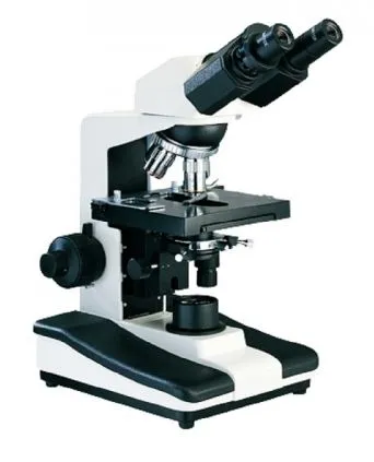
DARK FIELD MICROSCOPE SPECIAL
| Eyepiece |
Wide field PL10 × (F18mm) |
| Objective |
Plan achromatic objective:
4X/0.1, 10X/0.25, 40X/0.65(spr), 100X(spr.,oil).
Optional: 20X |
| Nosepiece |
Quadruple(Four-position). |
| Stage |
Stage size:160×140mm.
Moving range: 75mm ×50mm
Precision: 0.1mm |
| Focusing mechanism |
Coaxial coarse and fine focusing system.
Focusing stoper for protecting the lens and
specimen.
Tightness and height limitation of coarse
regulation are adjustable.
Fine adjustment accuracy :0.002mm. |
| Illumination |
Transmission illumination system.
6V/20W halogen lamp |
| Condenser |
Dark field conderser. |
| Camera adapter |
C-mount. |
| Camera |
5mp |
| Carry case |
Padded Aluminum |
| Power supply |
AC100-240V, 50/60Hz |
| |
|
|
|

|
Live
Blood Darkfield Microscope
With
hd camera
| Magnification |
40X - 1000X. |
| Application |
For living blood analysis.
For dry blood analysis. |
| Signal out put |
The image system support Monitor or TV with
AV interface.
With a video processor, it can connect to computer
with software for
image capturing and video recording. |
| View Tube |
Trinocular head, 30 ° incling, 360°rotating.
Diopter adjustable. Iinterpupillary distance
adjustable. |
| Eyepiece |
WF10X / 18mm. |
| Objective |
Plan Achromatic objective:
4X/0.1, 10X/0.25, 40X/0.65(Spr), 100X/1.25
(Spr. Oil). |
| Nosepiece |
Quadruple reversed angle nosepiece, revolver
with rotation on ball
bearings. |
| Stage |
Mechanical double layer stage, platinum with
overhang, dimensions
140x140 mm. Moving range: 75mm ×50mm.
Vernier scale on both axes of movement with
an accuracy of 0.1mm |
| Focusing |
Coaxial positioning system. Movement by roller
guider(Roller and pinion).
Rubber grip around fine and coarse adjust knob.
Tension adjustment of coarse regulation knob.
Upper pper limit stoper for protecting the
lens and sample. |
| Illumination |
3W LED cold light.
|
| Condenser |
Removable darkfield condenser, NA1.25 with
aperture iris diaphragm. |
| Video 1 |
Live Blood Watch
the Video |
| Video 2 |
Connect
to TV or computor screenWatch the video |
| |
Easy
to use with great Examples so you can analyze
and interpret your blood slide |
| |
 Full
Instructions Full
Instructions
Cetificate:
CE, Rohs. |
It
should be noted that Live Blood Analysis is
not a diagnostic procedure. This
method was designed as a screening test to
take the
guesswork out of selecting the appropriate
supplements for the individual patient.
|