At a recent evening lecture at the California Institute
of Technology, a neurologist was explaining the ins and
outs of new brain-imaging technology to an audience composed
of Caltech professors, students, and members of the general
public. The audience was rather quiet, lulled by the technical
tone of the lecture. But when the neurologist mentioned
in passing that the disease afflicting one of his patients
was caused by a brain parasite, the whole room sat up and
made a collective noise of disgust and alarm. Brain
parasites!
But, in fact, parasites infect us all the time. They live in our bodies,
even in our cells, and most of the time we do not even know that they
are there. The brain can provide a pleasant, nurturing environment
for parasites, because it has structures that prevent many of the immune
system’s cells from entering, at least in the early stages of
infection. Add to that plenty of oxygen and nutrients, and the brain
seems like a rather nice place to live.
Despite its seemingly idyllic home, a brain parasite’s life does
have its hardships. To begin with, the parasite has to find a way into
the brain. Invasion of any organ is difficult, but the brain is an especially
tough nut to crack due to a protective barrier between the bloodstream
and brain fluid, called the blood-brain barrier. This barrier is made
up of cells that make a tight seal along any blood vessels so that most
stuff from the bloodstream (including brain parasites) can’t leak
into the brain. If the parasite does manage to successfully enter the
brain, it then has to deal with the attack of the immune system. The
cells of the immune system act together to rid the body of any foreign
organisms. In humans, the immune system is highly organized and efficient;
parasites’ evasion mechanisms have evolved to be good enough to
thwart the immune system, at least for a little while. Unfortunately,
the most effective parasites are the ones we really have to worry about.
In fact, millions of people worldwide are infected by these efficacious
brain parasites. Many of these brain parasites cause debilitating conditions
and sometimes even death. So, in addition to being interesting biologically,
brain parasites are also important in the context of human disease.
Two parasites with disease-causing capabilities are the pork tapeworm,
Taenia solium, and the amoeba Naegleria fowleri. In addition to their
medical importance, these two organisms illustrate the many ways that
brain parasites are able to affect their hosts through their methods
of invasion and survival.
Tapeworm: From Pork Chops
to the Brain
The pork tapeworm is one of the most common disease-causing
brain parasites. This parasite infects over 50 million
people worldwide, and is the leading cause of brain seizures.
It is usually contracted from eating undercooked pork,
and once in the gut, it attaches to the intestine, and
then grows to be several feet long. Under certain circumstances,
these worms can also invade the brain, where thankfully
they don’t grow to be quite so large.
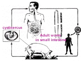
Why does the worm sometimes attach to the intestine
but at other times travel to the brain? It all depends
on what stage of its life cycle the worm is in when it
is swallowed. In its larval stage, the worm will hook
onto the intestine; however, if eggs are swallowed, they
hatch in the stomach. From there the larvae can enter
the bloodstream and eventually travel to the brain. But
in order to reach the brain from the bloodstream, the
larvae must traverse the blood-brain barrier. Unfortunately,
researchers still don’t know exactly how this happens.
Many scientists think that the larvae can release enzymes
that are able to dissolve a small portion of the blood-brain
barrier to allow the parasite to get through into the
brain.
Bob
Beck found Parasites can be illiminated
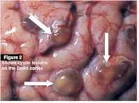
Once the larvae reach the brain, they cause a disease
called neurocysticercosis, by attaching to either the
brain tissue itself, or to cavities through which brain
fluid flows. (Brain fluid carries nutrients and waste
to and from the brain, and acts as a cushion to protect
the brain against physical impact.) Once attached, the
larvae develop into cyst-like structures. The location
of the cysts determines the symptoms exhibited by the
host. If the larvae attach to the brain tissue, then
the host often experiences seizures. This occurs partly
because the presence of the larvae causes the activity
of the brain to become wild and uncontrolled, thereby
causing a seizure. On the other hand, if the larvae attach
to the brain-fluid cavities, the host experiences headaches,
nausea, dizziness, and altered mental states in addition
to seizures. These additional symptoms occur because
the flow of the brain fluid is blocked by the larvae.
Often, the presence of the larvae also causes the lining
of the brain-fluid cavities to become inflamed, further
constricting the flow of the brain fluid. Since the cavities
are a closed system, blockage of the cavities exerts
pressure on the brain. This increased cranial pressure
forces the heart to pump harder in order to deliver blood
to the brain area, increasing the pressure on the brain
even more. If the condition is not treated, the heart
eventually cannot pump enough blood to the brain, neurons
begin to die off, and major brain damage occurs.
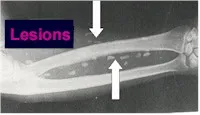
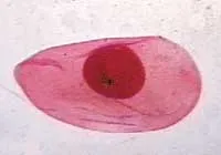 |
A pork tapeworm (Taenia solium)
cysticercus, the form in which the tapeworm is
found in an infected brain. (Colorized image
by P. W. Pappas and S. M. Wardrop, courtesy of
P. W. Pappas, Ohio State University.) |

Worm
on the Brain
Woman Recuperating After Doctors Remove Parasite
Which Parasites
Can Infect The Brain?
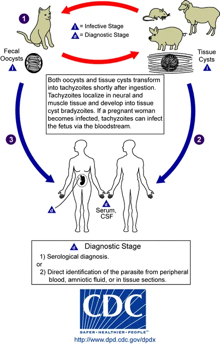
There are two known brain parasites, the pork tapeworm, Taenia
solium, and the amoeba Naegleria fowleri.
Taenia solium: The pig tapeworm, Taenia
solium, is responsible for the condition known
as neurocysticercosis, the most common brain
parasitic infection. Neurocysticercosis affects
more than fifty million people all over the
world, and it is the leading cause of brain
seizures. This disease develops when larvae
from Taenia solium enter the body via the ingestion
of diseased pork meat. Once inside the body,
the tapeworm migrates to the small intestine
and remains there until it reaches maturity.
From here the parasite makes its way to the
brain where it attaches either to the brain
tissues or to the cavities within the brain.
The parasite will then form cystic lesions
that can also affect the eyes, muscles and
spinal cord. The exact location of the cysts
will determine the symptoms of the disease.
Brain parasites can interrupt the normal activity
of the brain and cause brain seizures. On the
other hand, parasites that attach to the brain-fluid
cavities will cause symptoms such as headaches,
nausea, and dizziness, as well as brain seizures.
These additional symptoms may occur because
the parasite is interrupting the normal flow
of brain fluid within the brain. Over time,
this blockage of fluid may cause pressure to
build up that can lead to permanent brain damage.
Naegleria fowleri: Unlike the pork tapeworm,
Naegleria fowleri brain parasites have only
infected about 175 people in the world; therefore
it is not as easily known or understood. This
brain parasite causes a condition called primary
amoebic meningo-cephalitis. Of the 175 cases
of this disease that have been reported, only
six patients have survived. For this reason,
scientists are eagerly searching for more answers
as to these particular brain parasites can
be treated.
Naegleria fowleri is an amoeba that is commonly
found in the wild, especially in warm freshwater
lakes and ponds. It can also survive in heated
swimming pools. This parasite can infect a
human host that is swimming in contaminated
waters by attaching to the inside of its host's
nose and then traveling up the nose and into
the brain. Once in the area of the brain, the
amoeba releases an enzyme that allows it dissolve
the host's tissues, and enter the tissues of
the brain. Naegleria fowleri can then into
feast on the valuable nutrients with the neurons
of the brain. This is why this particular parasite
causes such rapid death.
In addition to the brain damage caused by
destroying the brain's neurons, the presence
this parasite can also cause inflammation of
the tissues within the brain. This inflammation
can lead to additional brain damage and even
death.
|
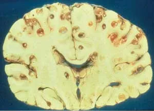
T. solium cysticerci in the brain of a nine-year-old
girl who died during cerebrospinal fluid extraction
to diagnose her headaches. This was in the 1970s—if
it had happened 10 years later, noninvasive computerized
tomography would have given an accurate diagnosis,
and the parasites could have been killed . (Image courtesy
of Dr. Ana Flisser, National Autonomous University
of Mexico.)
colloidal silver acts as
a "second immune
system" according to Bob Beck. It has been
shown in numerous studies to be the only substance
known to eliminate hundreds of viruses, bacteria,
fungus, etc., more than any modern antibiotic or "miracle
drug" yet developed by the pharmaceutical
companies.
More Here |
It is interesting to note that some of
these symptoms, such as seizures, are caused not only
by the presence of the brain parasites, but also by the
immune system. In general, parasites do not want to be
detected by the immune system, because then they will
most likely be eaten and killed. They try to do everything
they can to avoid eliciting a strong immune response.
Parasites also don’t want to do anything that can
kill the host. If the host dies, then the parasites die
too. For this reason, people can have parasites for years
and not show any symptoms at all. But then, as the larval
defenses break down, the host immune system is able to
have a greater effect, and the symptoms become more obvious.
What does the host immune system do to defend against
the parasites, and why do its actions elicit harmful
effects on its own body?
Defending the Body from
Invaders
The main function of the immune system is to make sure that any foreign
object in the body is destroyed, including brain parasites. Many of
the symptoms arising from brain parasite infection are due to the interactions
between the immune system and the parasite. There are two main methods
by which the immune system tries to rid the brain of the parasite.
First, certain cells of the immune system make antibodies specifically
against the parasite. Antibodies are molecules that can attach to a
foreign organism and act like a signal flare, telling the rest of the
immune cells that this organism is foreign and should be destroyed.
There are also other immune cells, called phagocytes, which travel
around the body eating anything that isn’t recognized as belonging
to that body. These cells are much more effective at destroying germs
that are labeled by antibodies.
Second, there are proteins in the body that are able to recognize some
general characteristics of many germs. These proteins make up the complement
system. The complement proteins are able to attach to the germ and
also act as signal flares to attract other immune cells that can destroy
the germ. However, these proteins are sometimes also able to kill the
germ themselves by forming a structure on the surface that can cut
the germ open.
Bob Beck Protocol
Packages |
|
|
|
|
| |
Universal detoxification is accomplished by
oxidation of dead and neutralized pathogens, without
the need for colonics, heat, hot tubs, exercise,
liver and kidney flushing, herbs or other modalities.
While these treatments have their uses, Bob Beck
feels they are not necessary if ozonated water
is used daily with the other three protocols he
recommends.
More
Here |
Why the
Immune System Can’t “See” Tapeworm
Cysts
The interaction between the immune system and the cysts is quite amazing;
it is a great example of how evolution can produce two complementary
systems. The immune system is seeking to find and destroy the parasite,
while the parasite is attempting to stay hidden and alive. One way
that the cysts are able to “hide” from the immune system
is by degrading the antibodies that attach to them. There is some evidence
that the antibodies are used as a food source, and that the cysts are
able to coax the immune system to make more antibodies. The cysts can
even disguise themselves as part of the host’s body by displaying
proteins on their surfaces that identify them as part of the host—much
as Wile E. Coyote hides from Sam Sheepdog in a herd of sheep by wearing
a sheepskin. Finally, the location of the cysts is itself conducive
to escaping detection by the immune system. The brain is not easily
accessible to the cells of the immune system due to the presence of
the blood-brain barrier, and so the parasites are partially protected
from random encounters with the body’s defenders. Only when the
immune response is in full swing can the immune cells enter the brain
in large numbers.
Besides hiding from the immune system, the tapeworm parasites are able
to prevent the immune cells from killing them by using several strategies.
For instance, the parasites are able to prevent the complement proteins
from attaching to their surfaces. The tapeworms can even release molecules
that act as decoys, tricking the killer proteins into leaving them
alone. The cysts also release other proteins that are able to protect
them from being eaten, although how exactly this is accomplished is
still unknown.
There is some evidence that these proteins are able
to prevent phagocytes from accurately targeting the cysts.
One of the ways that phagocytes are able to go to the
right place in the body during an infection is by following
a chemical trail. This trail is produced by other immune
cells at the site of infection. Some of the proteins
released by the cysts are able to obscure this chemical
trail so that the phagocytes become lost on their way
to the infection. Cysts are also thought to release a
second set of proteins that decreases the activity of
new phagocytes. These proteins affect another group of
immune cells that control the activity of new phagocytes;
these regulatory immune cells then decrease the number
of active phagocytes. Finally, a third set of proteins
released by the cysts is thought to be able to prevent
phagocytes from producing the proteins necessary to kill
the cysts.
This treatment disables microbes as they float
through the bloodstream. This is an important part
of the protocol.
Dr Bob Beck electrification devices are attached
directly to the bloodstream via the wrists, not
the palms of the hands, as with Dr
Clark's zapper. The Beck electrification device output is much
stronger and has been measured in the bloodstream,
using hypodermic probes.
The slower pulsation of Beck's device is said to "permit the
current to penetrate deeply".
Once the microbes are disabled, they are harmless
and the body should eventually excrete them.
More
info here |
Victory?
The cysts are very successful in evading the immune
system, but they gradually become more and more vulnerable
to attack. As the immune system response gains strength,
the most common symptoms of infection become more and
more obvious. At first, the parasites are simply unable
to hide from the immune cells, and cannot pretend to
be part of the host’s body anymore. Then the full
immune system response kicks in, and because the immune
cells are able to detect the parasites, the parasites
are doomed. More antibodies and complement proteins are
released, more phagocytes are born, and more blood and
immune cells rush to the parasitic sites. The areas where
the parasites are located become swollen, which often
leads to seizures and compression of the surrounding
brain tissue. As the response progresses, the cysts are
replaced by scar tissue, and finally by calcium deposits.
(Calcium deposition often occurs in the body due to the
activity of bacteria living in the blood, rather than
as a direct effect of the immune system’s response.)
The scar tissue and calcium deposits are also known to
cause seizures. In addition, the immune response causes
irreparable brain damage to the areas of the brain around
the cyst as the phagocytes ingest the cells surrounding
the cysts, which also contributes to the seizures.
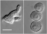 |
Naegleria fowleri in the amoeboid
form, near right, and in the cyst form, far right.
The scale bar is 10 micrometers. Images courtesy
of Bret Robinson, Australian Water Quality Centre
and CRC for Water Quality Research. |
In fact, more harm than good often comes out of the
immune response to infection of the brain by tapeworms.
Against most pathogens, however, the immune response
is actually beneficial to the body. Foreign organisms
often cause lots of damage, and it is important that
they be destroyed as quickly and efficiently as possible.
Furthermore, the immune system response is generally
the same regardless of the identity of the foreign invader;
and in most circumstances, the immune response does not
have negative effects. Overall, the immune system is
actually highly effective at defending the body from
foreign organisms.
Of course, the effectiveness of the immune system is largely dependent
on the ability of the body to mobilize its defenses. Some parasites
act so quickly that the immune system is unable to react before the
infection becomes fatal. One such brain parasite is Naegleria fowleri,
a water-borne amoeba.
Danger in the Waters
If you’ve never heard of Naegleria fowleri, don’t
be surprised. Unlike the pork tapeworm, N. fowleri has
only infected about 175 people in the world, causing
a disease called primary amoebic meningo-cephalitis.
But out of those 175 people, only six have survived,
giving a mortality rate of 97 percent. For this reason,
it is quite an important parasite to study, as there
are no current treatments that have proven effective
against it.
Fortunately, natural infection by the parasite is very rare, although
N. fowleri is ubiquitous in the wild. It lives mostly in warm freshwater
lakes and ponds, but can even thrive in heated swimming pools. Furthermore,
N. fowleri is actually a free-living organism, which means that it
can survive without a host. This explains why N. fowleri attacks are
so rapidly fatal—since hosts are not necessary to its survival,
the parasite does not have to take pains to avoid killing them.
Part of the reason that N. fowleri can survive in such numbers and in
so many different places is because it is an amoeba. Amoebas are single-celled
creatures that resemble sacks of fluid gelatin surrounded by a greasy
membrane. Because of their small size and few requisites for survival,
these organisms are found everywhere. In addition, the amoebas can
form cysts in harsh conditions like extreme cold; in this form, they
are protected against the environment.
Attack of the Amoebas
When an amoeba invades a person, it is normally in
its active, reproductive phase. Invasion occurs when
the amoeba attaches to the inside of its host’s
nose and then travels up the nose to the brain. The amoeba
follows the path laid out by the olfactory nerve, although
sometimes it can also use the bloodstream. Several enzymes
released by the amoeba are able to dissolve the host’s
tissues, giving access to the brain. Once in the brain,
the amoeba causes damage by actually eating the nerve
cells. As you can imagine, this is very harmful to the
host, and is the main reason why infection by N. fowleri
causes such rapid death. The amoeba is able to eat neurons
because it has surface proteins that allow it to cut
a hole in the covering of the cell. The contents of the
neuron leak out, and the amoeba can feed on the nutrients
it contains. The amoeba even has proteins on its surface
that tell it where the best food sources are. These proteins
are able to sense the presence of certain nutrients,
and then send signals to the rest of the cell indicating
in which direction the amoeba should move to eat those
nutrients. Finally, there are other proteins on the amoeba’s
surface that direct it to the most vulnerable areas of
a neuron.
In addition to causing direct brain damage by ingesting neurons, the
presence of N. fowleri amoebas can cause inflammation of the brain-fluid
cavity linings. Similarly to infection by tapeworm, blocking the brain
fluid can cause increased pressure on the brain. However, this effect
is usually only secondary to the much more destructive digesting action
of the amoebas.
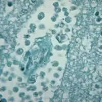 |
Brain tissue infected by Naegleria
fowleri. The dark dots are the amoebas. Notice
the empty space around the dots; this space used
to be tissue before the amoebas digested it.
Image provided by the Division of Parasitic Diseases,
Centers for Disease Control and Prevention. |
Fighting the Invader
The immune system, however, is not completely idle
while this invasion and destruction is occurring, although
for the most part its efforts are in vain. The amoebas
use several strategies to stave off the immune cells.
Many of these strategies are similar to those used by
tapeworm cysts. For example, the amoebas are able to
internalize antibodies on their surfaces, although they
don’t need these antibodies as a food source. Other
proteins on the amoeba’s surface prevent the attachment
of complement proteins. If the complement proteins are
able to bypass these surface proteins, the amoeba is
able to collect them in one area of its membrane. Afterwards,
the amoeba can shed that piece of the membrane. The shed
membrane acts as a decoy, attracting more complement
proteins that would otherwise attack the amoeba.
The concept has been revealed in many revolutionary
patents and research papers over the past 100
years (going back to 1890), but these breakthroughs
were typically lost, suppressed, ridiculed by
mainstream medicine, etc. Blood electrification
takes 2 hours daily for about four weeks to get
significant results.
More
info Here |
Why are these strategies effective in shielding the
amoebas, but not tapeworms, from the immune system? The
reason is that an amoebal infection is rapidly fatal.
The immune system does not have time to fully mobilize
its immune cell armies before the brain damage is so
extreme that the organism dies. Since these amoebas don’t
need the host to survive, it’s not a big deal if
they kill him or her off. Tapeworms, however, die when
the host does, and so they try very hard to keep from
being detected by the immune system. And in fact, they
do a fairly good job at that, since most tapeworm infections
aren’t noticeable until many years after the tapeworms
get into the brain. The immune system is only able to
have a big effect on the infection when the tapeworms
start to die, often from old age.
Parasite Evolution
These two parasites offer only an inkling of the many
organisms that can infect the human brain. While the
two seem to differ greatly, the molecular weapons they
use for defense and invasion are really very similar.
For instance, there is evidence that both parasites use
enzymes to penetrate the blood-brain barrier, and both
use a decoy strategy to deflect the attention of the
immune system. This similarity results from evolution,
which has slowly altered these parasites so that they
are as effective as possible at survival. As new treatments
and cures of brain-parasite-related diseases become available,
it will be interesting (as well as medically useful)
to see how the strategies of these parasites change.
BOB
BECK PROTOCOL
Bob Beck Protocol
Packages |
|
|
|
|
By
Andrea Manzo
Andrea Manzo is a senior majoring in biology. She decided to find out
more about brain parasites after attending the 2002 Biology Forum, “Gray
Matters: |